4P24

pore forming toxin


Experimental Data Snapshot

  • Method: X-RAY DIFFRACTION
  • Resolution: 3.10 Å
  • R-Value Free: 0.244 
  • R-Value Work: 0.219 
  • R-Value Observed: 0.220 

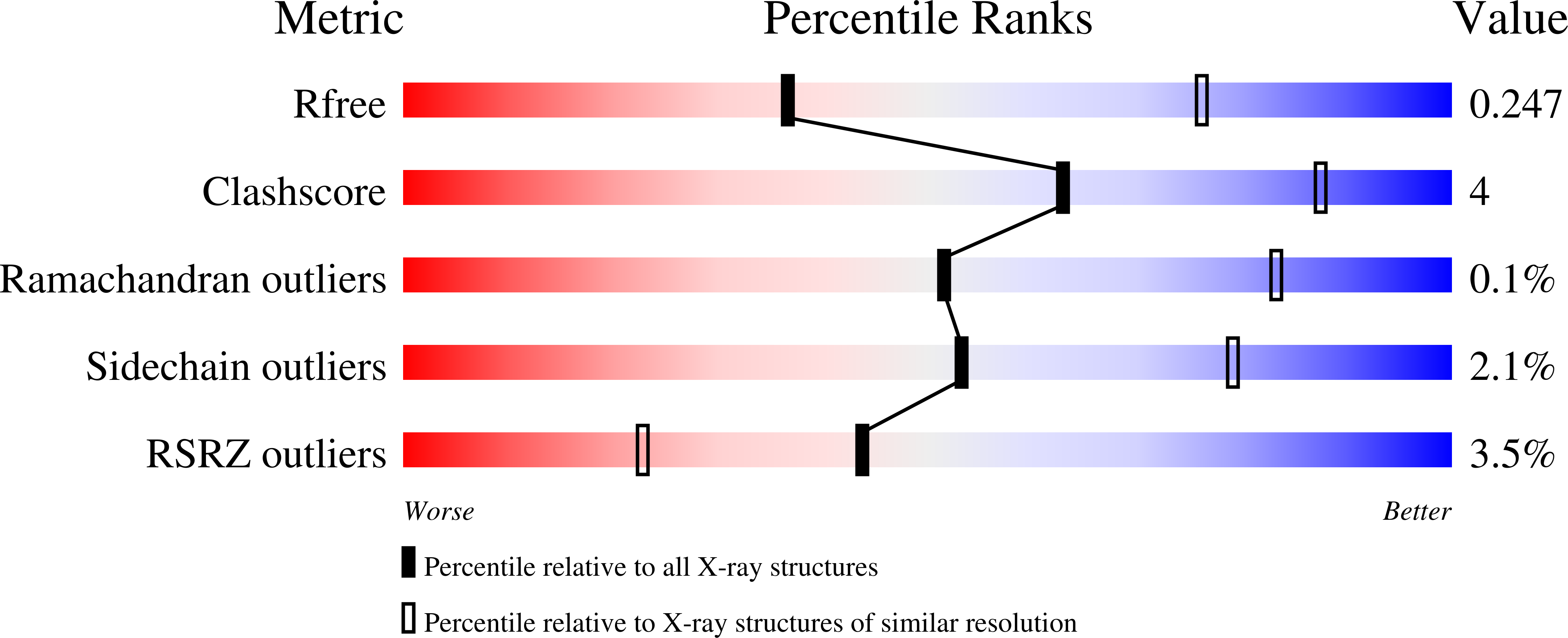
wwPDB Validation   3D Report Full Report


This is version 1.2 of the entry. See complete history


Literature

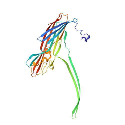
Structural basis for pore-forming mechanism of staphylococcal alpha-hemolysin.

Sugawara, T.Yamashita, D.Kato, K.Peng, Z.Ueda, J.Kaneko, J.Kamio, Y.Tanaka, Y.Yao, M.

(2015) Toxicon 108: 226-231

  • DOI: https://doi.org/10.1016/j.toxicon.2015.09.033
  • Primary Citation of Related Structures:  
    4P24, 4YHD

  • PubMed Abstract: 

    Staphylococcal α-hemolysin (α-HL) is a β-barrel pore-forming toxin (PFT) expressed by Staphylococcus aureus. α-HL is secreted as a water-soluble monomeric protein, which binds to target membranes and forms membrane-inserted heptameric pores. To explore the pore-forming mechanism of α-HL in detail, we determined the crystal structure of the α-HL monomer and prepore using H35A mutant and W179A/R200A mutant, respectively. Although the overall structure of the monomer was similar to that of other staphylococcal PFTs, a marked difference was observed in the N-terminal amino latch, which bent toward the prestem. Moreover, the prestem was fastened by the cap domain with a key hydrogen bond between Asp45 and Tyr118. Prepore structure showed that the transmembrane region is roughly formed with flexibility, although the upper half of the β-barrel is formed appropriately. Structure comparison among monomer, prepore and pore revealed a series of motions, in which the N-terminal amino latch released upon oligomerization destroys its own key hydrogen bond between Asp45-Tyr118. This action initiated the protrusion of the prestem. Y118F mutant and the N-terminal truncated mutant markedly decreased in the hemolytic activity, indicating the importance of the key hydrogen bond and the N-terminal amino latch on the pore formation. Based on these observations, we proposed a dynamic molecular mechanism of pore formation for α-HL.


  • Organizational Affiliation

    Graduate School of Life Science, Hokkaido University, Sapporo, 060-0810, Japan.


Macromolecules
Find similar proteins by:  (by identity cutoff)  |  3D Structure
Entity ID: 1
MoleculeChains Sequence LengthOrganismDetailsImage
Alpha-hemolysin
A, B, C, D, E
A, B, C, D, E, F, G
302Staphylococcus aureus subsp. aureus Mu50Mutation(s): 2 
Gene Names: SAV1163
UniProt
Find proteins for A0A0J9X1Z2 (Staphylococcus aureus (strain Mu50 / ATCC 700699))
Explore A0A0J9X1Z2 
Go to UniProtKB:  A0A0J9X1Z2
Entity Groups  
Sequence Clusters30% Identity50% Identity70% Identity90% Identity95% Identity100% Identity
UniProt GroupA0A0J9X1Z2
Sequence Annotations
Expand
  • Reference Sequence
Small Molecules
Ligands 1 Unique
IDChains Name / Formula / InChI Key2D Diagram3D Interactions
MPD
Query on MPD

Download Ideal Coordinates CCD File 
H [auth A]
I [auth A]
J [auth A]
K [auth B]
L [auth C]
H [auth A],
I [auth A],
J [auth A],
K [auth B],
L [auth C],
M [auth C],
N [auth D],
O [auth D],
P [auth E],
Q [auth F],
R [auth F],
S [auth G],
T [auth G]
(4S)-2-METHYL-2,4-PENTANEDIOL
C6 H14 O2
SVTBMSDMJJWYQN-YFKPBYRVSA-N
Experimental Data & Validation

Experimental Data

  • Method: X-RAY DIFFRACTION
  • Resolution: 3.10 Å
  • R-Value Free: 0.244 
  • R-Value Work: 0.219 
  • R-Value Observed: 0.220 
  • Space Group: P 41 21 2
Unit Cell:
Length ( Å )Angle ( ˚ )
a = 170.06α = 90
b = 170.06β = 90
c = 202.86γ = 90
Software Package:
Software NamePurpose
PHENIXrefinement

Structure Validation

View Full Validation Report


Entry History 

Deposition Data

Revision History  (Full details and data files)

  • Version 1.0: 2015-03-11
    Type: Initial release
  • Version 1.1: 2018-03-21
    Changes: Advisory, Data collection, Database references, Derived calculations, Other, Source and taxonomy, Structure summary
  • Version 1.2: 2023-12-27
    Changes: Data collection, Database references, Derived calculations, Refinement description