4OTY

Crystal structure of lumiracoxib bound to the apo-mouse-cyclooxygenase-2


Experimental Data Snapshot

  • Method: X-RAY DIFFRACTION
  • Resolution: 2.35 Å
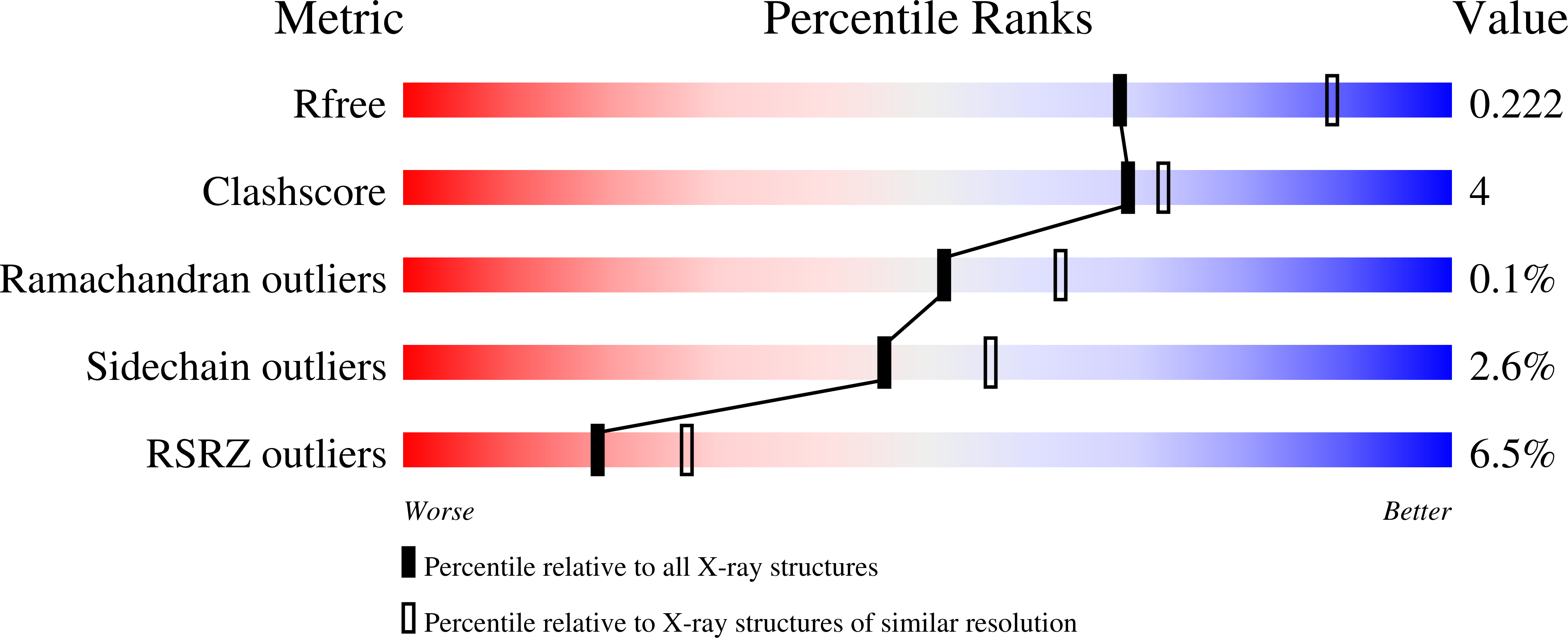
  • R-Value Free: 0.221 
  • R-Value Work: 0.180 
  • R-Value Observed: 0.181 

wwPDB Validation   3D Report Full Report


Ligand Structure Quality Assessment 


This is version 2.1 of the entry. See complete history


Literature

Exploring the molecular determinants of substrate-selective inhibition of cyclooxygenase-2 by lumiracoxib.

Windsor, M.A.Valk, P.L.Xu, S.Banerjee, S.Marnett, L.J.

(2013) Bioorg Med Chem Lett 23: 5860-5864

  • DOI: https://doi.org/10.1016/j.bmcl.2013.08.097
  • Primary Citation of Related Structures:  
    4OTY

  • PubMed Abstract: 

    Lumiracoxib is a substrate-selective inhibitor of endocannabinoid oxygenation by cyclooxygenase-2 (COX-2). We assayed a series of lumiracoxib derivatives to identify the structural determinants of substrate-selective inhibition. The hydrogen-bonding potential of the substituents at the ortho positions of the aniline ring dictated the potency and substrate selectivity of the inhibitors. The presence of a 5'-methyl group on the phenylacetic acid ring increased the potency of molecules with a single ortho substituent. Des-fluorolumiracoxib (2) was the most potent and selective inhibitor of endocannabinoid oxygenation. The positioning of critical substituents in the binding site was identified from a 2.35Å crystal structure of lumiracoxib bound to COX-2.


  • Organizational Affiliation

    A.B. Hancock Jr. Memorial Laboratory for Cancer Research, Department of Biochemistry, Department of Chemistry, Department of Pharmacology, Vanderbilt Institute of Chemical Biology, Center in Molecular Toxicology, and Vanderbilt-Ingram Cancer Center, Vanderbilt University School of Medicine, Nashville, TN, United States.


Macromolecules
Find similar proteins by:  (by identity cutoff)  |  3D Structure
Entity ID: 1
MoleculeChains Sequence LengthOrganismDetailsImage
Prostaglandin G/H synthase 2
A, B
587Mus musculusMutation(s): 0 
Gene Names: Ptgs2Cox-2Cox2Pghs-bTis10
EC: 1.14.99.1
UniProt
Find proteins for Q05769 (Mus musculus)
Explore Q05769 
Go to UniProtKB:  Q05769
Entity Groups  
Sequence Clusters30% Identity50% Identity70% Identity90% Identity95% Identity100% Identity
UniProt GroupQ05769
Sequence Annotations
Expand
  • Reference Sequence
Oligosaccharides

Help

Entity ID: 2
MoleculeChains Length2D Diagram Glycosylation3D Interactions
2-acetamido-2-deoxy-beta-D-glucopyranose-(1-4)-2-acetamido-2-deoxy-beta-D-glucopyranose
C, D
2N-Glycosylation
Glycosylation Resources
GlyTouCan:  G42666HT
GlyCosmos:  G42666HT
GlyGen:  G42666HT
Small Molecules
Binding Affinity Annotations 
IDSourceBinding Affinity
LUR BindingDB:  4OTY Ki: 320 (nM) from 1 assay(s)
IC50: min: 7, max: 1.00e+4 (nM) from 7 assay(s)
Binding MOAD:  4OTY IC50: 40 (nM) from 1 assay(s)
Experimental Data & Validation

Experimental Data

  • Method: X-RAY DIFFRACTION
  • Resolution: 2.35 Å
  • R-Value Free: 0.221 
  • R-Value Work: 0.180 
  • R-Value Observed: 0.181 
  • Space Group: I 2 2 2
Unit Cell:
Length ( Å )Angle ( ˚ )
a = 122.663α = 90
b = 133.173β = 90
c = 181.546γ = 90
Software Package:
Software NamePurpose
HKL-2000data collection
PHASERphasing
PHENIXrefinement
XDSdata reduction
SCALEPACKdata scaling

Structure Validation

View Full Validation Report


Ligand Structure Quality Assessment 


Entry History 

Deposition Data

Revision History  (Full details and data files)

  • Version 1.0: 2014-02-26
    Type: Initial release
  • Version 2.0: 2020-07-29
    Type: Remediation
    Reason: Carbohydrate remediation
    Changes: Atomic model, Data collection, Derived calculations, Structure summary
  • Version 2.1: 2023-09-20
    Changes: Data collection, Database references, Refinement description, Structure summary