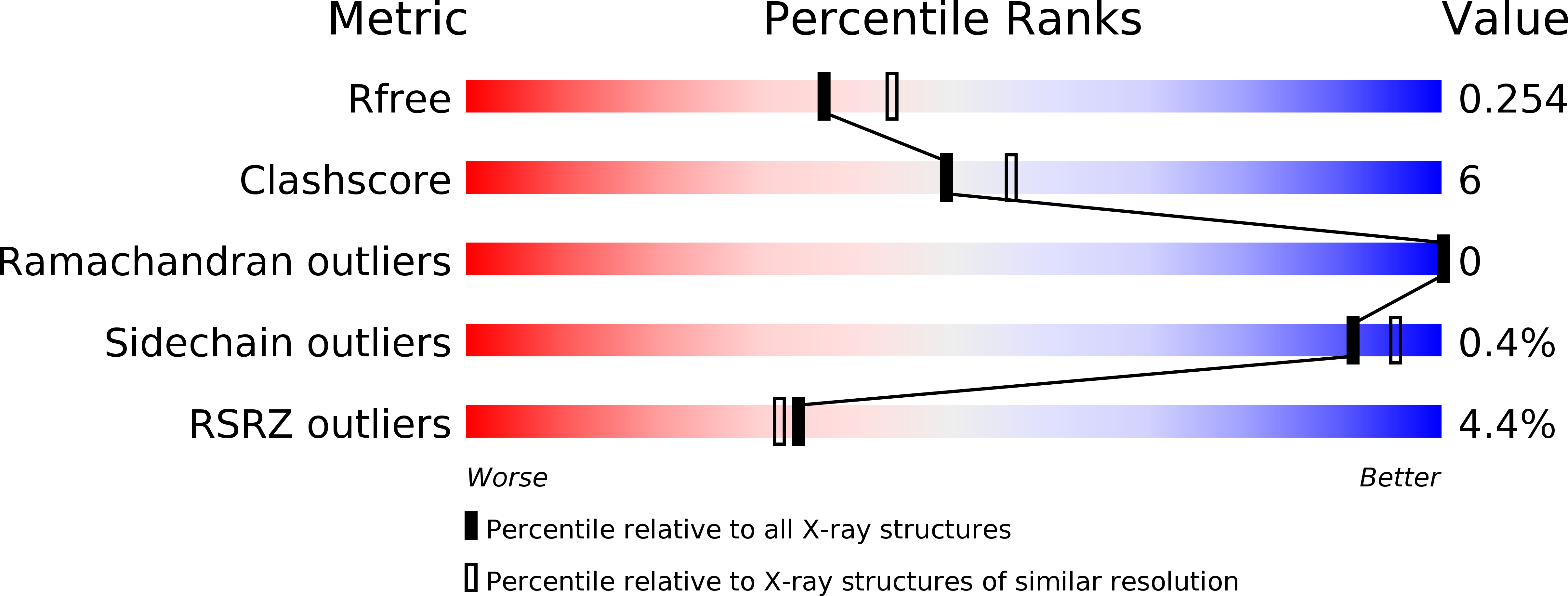
4OQW

Crystal structure of mCardinal far-red fluorescent protein


Experimental Data Snapshot

  • Method: X-RAY DIFFRACTION
  • Resolution: 2.21 Å
  • R-Value Free: 0.251 
  • R-Value Work: 0.223 
  • R-Value Observed: 0.225 

wwPDB Validation   3D Report Full Report


This is version 1.1 of the entry. See complete history


Literature

Non-invasive intravital imaging of cellular differentiation with a bright red-excitable fluorescent protein.

Chu, J.Haynes, R.D.Corbel, S.Y.Li, P.Gonzalez-Gonzalez, E.Burg, J.S.Ataie, N.J.Lam, A.J.Cranfill, P.J.Baird, M.A.Davidson, M.W.Ng, H.L.Garcia, K.C.Contag, C.H.Shen, K.Blau, H.M.Lin, M.Z.

(2014) Nat Methods 11: 572-578

  • DOI: https://doi.org/10.1038/nmeth.2888
  • Primary Citation of Related Structures:  
    4OJ0, 4OQW

  • PubMed Abstract: 

    A method for non-invasive visualization of genetically labeled cells in animal disease models with micrometer-level resolution would greatly facilitate development of cell-based therapies. Imaging of fluorescent proteins (FPs) using red excitation light in the 'optical window' above 600 nm is one potential method for visualizing implanted cells. However, previous efforts to engineer FPs with peak excitation beyond 600 nm have resulted in undesirable reductions in brightness. Here we report three new red-excitable monomeric FPs obtained by structure-guided mutagenesis of mNeptune. Two of these, mNeptune2 and mNeptune2.5, demonstrate improved maturation and brighter fluorescence than mNeptune, whereas the third, mCardinal, has a red-shifted excitation spectrum without reduction in brightness. We show that mCardinal can be used to non-invasively and longitudinally visualize the differentiation of myoblasts into myocytes in living mice with high anatomical detail.


  • Organizational Affiliation

    1] Department of Bioengineering, Stanford University, Stanford, California, USA. [2] Department of Pediatrics, Stanford University School of Medicine, Stanford, California, USA.


Macromolecules
Find similar proteins by:  (by identity cutoff)  |  3D Structure
Entity ID: 1
MoleculeChains Sequence LengthOrganismDetailsImage
Fluorescent protein FP480
A, B, C, D, E
A, B, C, D, E, F, G, H
228Entacmaea quadricolorMutation(s): 1 
UniProt
Find proteins for D0VX33 (Entacmaea quadricolor)
Explore D0VX33 
Go to UniProtKB:  D0VX33
Entity Groups  
Sequence Clusters30% Identity50% Identity70% Identity90% Identity95% Identity100% Identity
UniProt GroupD0VX33
Sequence Annotations
Expand
  • Reference Sequence
Small Molecules
Modified Residues  2 Unique
IDChains TypeFormula2D DiagramParent
MLY
Query on MLY
A, B, C, D, E
A, B, C, D, E, F, G, H
L-PEPTIDE LINKINGC8 H18 N2 O2LYS
NRQ
Query on NRQ
A, B, C, D, E
A, B, C, D, E, F, G, H
L-PEPTIDE LINKINGC16 H17 N3 O4 SMET, TYR, GLY
Experimental Data & Validation

Experimental Data

  • Method: X-RAY DIFFRACTION
  • Resolution: 2.21 Å
  • R-Value Free: 0.251 
  • R-Value Work: 0.223 
  • R-Value Observed: 0.225 
  • Space Group: P 1 21 1
Unit Cell:
Length ( Å )Angle ( ˚ )
a = 53.2α = 90
b = 136.7β = 90.1
c = 167.6γ = 90
Software Package:
Software NamePurpose
XSCALEdata scaling
PHASERphasing
PHENIXrefinement
PDB_EXTRACTdata extraction
XDSdata scaling

Structure Validation

View Full Validation Report


Entry History 

Deposition Data

Revision History  (Full details and data files)

  • Version 1.0: 2014-03-12
    Type: Initial release
  • Version 1.1: 2014-05-14
    Changes: Database references