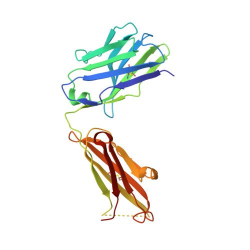
Water channel in the binding site of a high affinity anti-methotrexate antibody.
Gayda, S., Longenecker, K.L., Manoj, S., Judge, R.A., Saldana, S.C., Ruan, Q., Swift, K.M., Tetin, S.Y.(2014) Biochemistry 53: 3719-3726
- PubMed: 24832237
- DOI: https://doi.org/10.1021/bi5001382
- Primary Citation of Related Structures:
4OCX, 4OCY - PubMed Abstract:
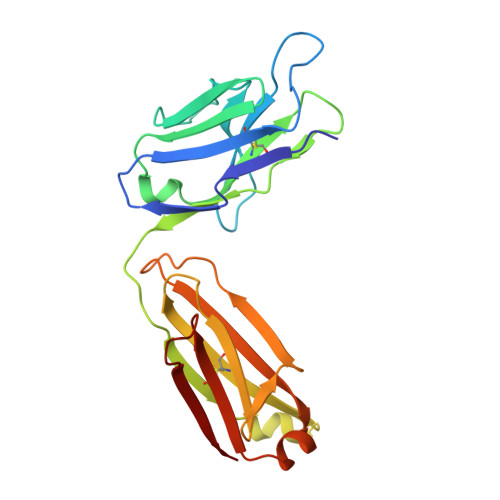
In the present study, we report the structure of the free and drug-bound Fab fragment of a high affinity anti-methotrexate antibody and perform a thermodynamic analysis of the binding process. The anti-methotrexate Fab fragment features a remarkably rigid tunnel-like binding site that extends into a water channel serving as a specialized route to move solvent out and into the site upon ligand binding and dissociation. This new finding in antibody structure-function relationships directly relates to the fast association (1 × 10⁷ M⁻¹ s⁻¹) and slow dissociation (4 × 10⁻⁵ s⁻¹) rates determined for mAb ADD056, resulting in a very strong binding with a K(D) ~ 3.6 pM at 20 °C. As follows from the X-ray data analysis, the methotrexate-antibody complex is stabilized by an extended network of hydrogen bonds and stacking interactions. The analysis also shows structural involvement of the CDR H3 in formation of the water channel revealing another important role of this hypervariable region. This suggests a new direction in natural affinity maturation and opens a new possibility in antibody engineering. Methotrexate is a widely used therapeutic agent for many malignant diseases and inflammatory disorders. Unfortunately, it may also interfere with central aspects of metabolism and thereby cause inevitable side effects. Therefore, methotrexate therapy requires careful monitoring of drug blood levels, which is traditionally done by immunoassays. An understanding of the structure-function properties of antibodies selected for drug monitoring substantiates the performance and robustness of such tests.
Organizational Affiliation:
Diagnostics Research, Abbott Diagnostics Division and ‡Structural Biology, Global Pharmaceutical Research and Development, Abbott Laboratories , Abbott Park, Illinois 60064, United States.