4O6G

Rv3902c from M. tuberculosis


Experimental Data Snapshot

  • Method: X-RAY DIFFRACTION
  • Resolution: 1.55 Å
  • R-Value Free: 0.177 
  • R-Value Work: 0.165 
  • R-Value Observed: 0.166 

wwPDB Validation   3D Report Full Report


This is version 1.3 of the entry. See complete history


Literature

1.55 angstrom resolution X-ray crystal structure of Rv3902c from Mycobacterium tuberculosis.

Reddy, B.G.Moates, D.B.Kim, H.B.Green, T.J.Kim, C.Y.Terwilliger, T.C.Delucas, L.J.

(2014) Acta Crystallogr Sect F Struct Biol Cryst Commun 70: 414-417

  • DOI: https://doi.org/10.1107/S2053230X14003793
  • Primary Citation of Related Structures:  
    4O6G

  • PubMed Abstract: 

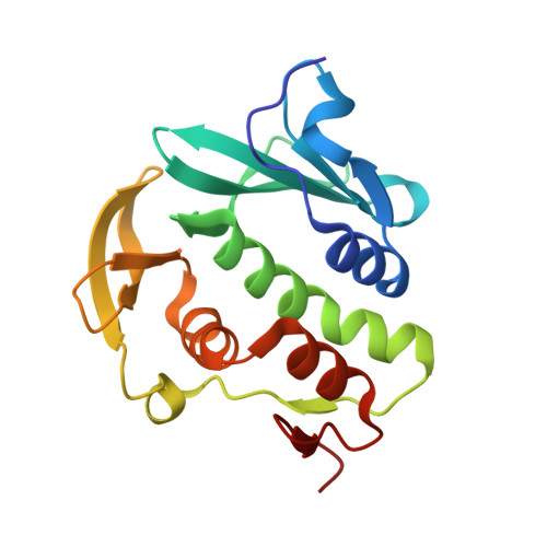
    The crystallographic structure of the Mycobacterium tuberculosis (TB) protein Rv3902c (176 residues; molecular mass of 19.8 kDa) was determined at 1.55 Å resolution. The function of Rv3902c is unknown, although several TB genes involved in bacterial pathogenesis are expressed from the operon containing the Rv3902c gene. The unique structural fold of Rv3902c contains two domains, each consisting of antiparallel β-sheets and α-helices, creating a hand-like binding motif with a small binding pocket in the palm. Structural homology searches reveal that Rv3902c has an overall structure similar to that of the Salmonella virulence-factor chaperone InvB, with an r.m.s.d. for main-chain atoms of 2.3 Å along an aligned domain.


  • Organizational Affiliation

    Department of Biochemistry and Molecular Genetics, University of Alabama at Birmingham, 1025 18th Street South, Birmingham, AL 35233, USA.


Macromolecules
Find similar proteins by:  (by identity cutoff)  |  3D Structure
Entity ID: 1
MoleculeChains Sequence LengthOrganismDetailsImage
Uncharacterized protein174Mycobacterium tuberculosisMutation(s): 0 
Gene Names: Mb3932cMT4020Rv3902c
UniProt
Find proteins for L7N519 (Mycobacterium tuberculosis (strain CDC 1551 / Oshkosh))
Explore L7N519 
Go to UniProtKB:  L7N519
Entity Groups  
Sequence Clusters30% Identity50% Identity70% Identity90% Identity95% Identity100% Identity
UniProt GroupL7N519
Sequence Annotations
Expand
  • Reference Sequence
Experimental Data & Validation

Experimental Data

  • Method: X-RAY DIFFRACTION
  • Resolution: 1.55 Å
  • R-Value Free: 0.177 
  • R-Value Work: 0.165 
  • R-Value Observed: 0.166 
  • Space Group: P 61
Unit Cell:
Length ( Å )Angle ( ˚ )
a = 91.819α = 90
b = 91.819β = 90
c = 54.231γ = 120
Software Package:
Software NamePurpose
SERGUIdata collection
PHENIXmodel building
PHENIXrefinement
MOSFLMdata reduction
Aimlessdata scaling
PHENIXphasing

Structure Validation

View Full Validation Report


Entry History 

Deposition Data

Revision History  (Full details and data files)

  • Version 1.0: 2014-03-05
    Type: Initial release
  • Version 1.1: 2014-04-16
    Changes: Database references
  • Version 1.2: 2024-02-28
    Changes: Data collection, Database references
  • Version 1.3: 2024-04-03
    Changes: Refinement description