4O27

Crystal structure of MST3-MO25 complex with WIF motif


Experimental Data Snapshot

  • Method: X-RAY DIFFRACTION
  • Resolution: 3.19 Å
  • R-Value Free: 0.260 
  • R-Value Work: 0.197 
  • R-Value Observed: 0.203 

wwPDB Validation   3D Report Full Report


This is version 1.1 of the entry. See complete history


Literature

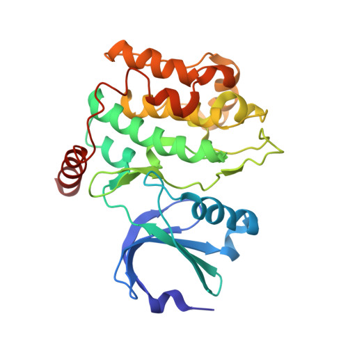
Structural insights into regulatory mechanisms of MO25-mediated kinase activation.

Hao, Q.Feng, M.Shi, Z.Li, C.Chen, M.Wang, W.Zhang, M.Jiao, S.Zhou, Z.

(2014) J Struct Biol 186: 224-233

  • DOI: https://doi.org/10.1016/j.jsb.2014.04.005
  • Primary Citation of Related Structures:  
    4NZW, 4O27

  • PubMed Abstract: 

    The tumor suppressor kinase LKB1 and germinal center kinases (GCKs) are key regulators of various cellular functions. The adaptor molecule MO25 not only recruits and activates LKB1 through the pseudokinase STRAD, but also may directly activate GCKs like MST3, MST4, STK25, OSR1 and SPAK. Targeting MO25 in a pathological setting has been recently studied in mouse. Yet the regulatory mechanism of MO25-mediated kinase activation is not fully understood. Here, our structural studies of MO25-related kinases reveal that MO25 binds to and activates GCK kinases or pseudokinase through a unified structural mechanism, featuring an active conformation of the αC helix and A-loop stabilized by MO25. Compared to GCKs that are directly activated by MO25-binding, activation of LKB1 has evolved additional layer of regulatory machinery, i.e., MO25 "activates" the pseudokinase STRAD, which in turn activates LKB1. Importantly, the structures of MO25α-STK25 and MO25α-MST3 determined in this work represent a transition/intermediate state and a fully activated state, respectively during the MO25-mediated kinase activating process.


  • Organizational Affiliation

    Institute of Biochemistry and Cell Biology, Shanghai Institutes for Biological Sciences, Chinese Academy of Sciences, Shanghai 200031, China.


Macromolecules
Find similar proteins by:  (by identity cutoff)  |  3D Structure
Entity ID: 1
MoleculeChains Sequence LengthOrganismDetailsImage
Calcium-binding protein 39325Homo sapiensMutation(s): 0 
Gene Names: CAB39CGI-66MO25
UniProt & NIH Common Fund Data Resources
Find proteins for Q9Y376 (Homo sapiens)
Explore Q9Y376 
Go to UniProtKB:  Q9Y376
PHAROS:  Q9Y376
GTEx:  ENSG00000135932 
Entity Groups  
Sequence Clusters30% Identity50% Identity70% Identity90% Identity95% Identity100% Identity
UniProt GroupQ9Y376
Sequence Annotations
Expand
  • Reference Sequence
Find similar proteins by:  (by identity cutoff)  |  3D Structure
Entity ID: 2
MoleculeChains Sequence LengthOrganismDetailsImage
Serine/threonine-protein kinase 24281Homo sapiensMutation(s): 1 
Gene Names: MST3STK24STK3
EC: 2.7.11.1
UniProt & NIH Common Fund Data Resources
Find proteins for Q9Y6E0 (Homo sapiens)
Explore Q9Y6E0 
Go to UniProtKB:  Q9Y6E0
PHAROS:  Q9Y6E0
GTEx:  ENSG00000102572 
Entity Groups  
Sequence Clusters30% Identity50% Identity70% Identity90% Identity95% Identity100% Identity
UniProt GroupQ9Y6E0
Sequence Annotations
Expand
  • Reference Sequence

Find similar proteins by:  Sequence   |   3D Structure  

Entity ID: 3
MoleculeChains Sequence LengthOrganismDetailsImage
5-mer peptide from serine/threonine-protein kinase 245N/AMutation(s): 1 
EC: 2.7.11.1
UniProt & NIH Common Fund Data Resources
Find proteins for Q9Y6E0 (Homo sapiens)
Explore Q9Y6E0 
Go to UniProtKB:  Q9Y6E0
PHAROS:  Q9Y6E0
GTEx:  ENSG00000102572 
Entity Groups  
UniProt GroupQ9Y6E0
Sequence Annotations
Expand
  • Reference Sequence
Small Molecules
Ligands 2 Unique
IDChains Name / Formula / InChI Key2D Diagram3D Interactions
ADP
Query on ADP

Download Ideal Coordinates CCD File 
D [auth B]ADENOSINE-5'-DIPHOSPHATE
C10 H15 N5 O10 P2
XTWYTFMLZFPYCI-KQYNXXCUSA-N
MN
Query on MN

Download Ideal Coordinates CCD File 
E [auth B]MANGANESE (II) ION
Mn
WAEMQWOKJMHJLA-UHFFFAOYSA-N
Experimental Data & Validation

Experimental Data

  • Method: X-RAY DIFFRACTION
  • Resolution: 3.19 Å
  • R-Value Free: 0.260 
  • R-Value Work: 0.197 
  • R-Value Observed: 0.203 
  • Space Group: P 43 21 2
Unit Cell:
Length ( Å )Angle ( ˚ )
a = 119.111α = 90
b = 119.111β = 90
c = 149.258γ = 90
Software Package:
Software NamePurpose
HKL-2000data collection
PHASERphasing
PHENIXrefinement
HKL-2000data reduction
HKL-2000data scaling

Structure Validation

View Full Validation Report


Entry History 

Deposition Data

Revision History  (Full details and data files)

  • Version 1.0: 2014-12-03
    Type: Initial release
  • Version 1.1: 2023-11-08
    Changes: Data collection, Database references, Derived calculations, Refinement description