4O0T

Back pocket flexibility provides group-II PAK selectivity for type 1 kinase inhibitors


Experimental Data Snapshot

  • Method: X-RAY DIFFRACTION
  • Resolution: 2.60 Å
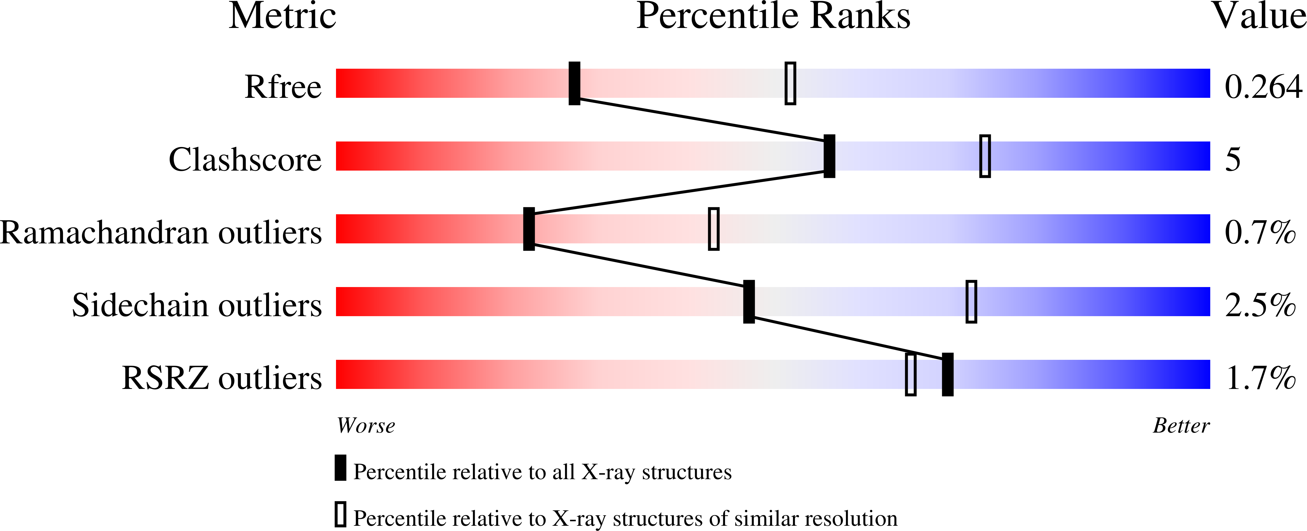
  • R-Value Free: 0.265 
  • R-Value Work: 0.214 
  • R-Value Observed: 0.217 

wwPDB Validation   3D Report Full Report


Ligand Structure Quality Assessment 


This is version 1.2 of the entry. See complete history


Literature

Back Pocket Flexibility Provides Group II p21-Activated Kinase (PAK) Selectivity for Type I 1/2 Kinase Inhibitors.

Staben, S.T.Feng, J.A.Lyle, K.Belvin, M.Boggs, J.Burch, J.D.Chua, C.C.Cui, H.Dipasquale, A.G.Friedman, L.S.Heise, C.Koeppen, H.Kotey, A.Mintzer, R.Oh, A.Roberts, D.A.Rouge, L.Rudolph, J.Tam, C.Wang, W.Xiao, Y.Young, A.Zhang, Y.Hoeflich, K.P.

(2014) J Med Chem 57: 1033-1045

  • DOI: https://doi.org/10.1021/jm401768t
  • Primary Citation of Related Structures:  
    4O0R, 4O0T, 4O0V, 4O0X, 4O0Y

  • PubMed Abstract: 

    Structure-based methods were used to design a potent and highly selective group II p21-activated kinase (PAK) inhibitor with a novel binding mode, compound 17. Hydrophobic interactions within a lipophilic pocket past the methionine gatekeeper of group II PAKs approached by these type I 1/2 binders were found to be important for improving potency. A structure-based hypothesis and strategy for achieving selectivity over group I PAKs, and the broad kinome, based on unique flexibility of this lipophilic pocket, is presented. A concentration-dependent decrease in tumor cell migration and invasion in two triple-negative breast cancer cell lines was observed with compound 17.


  • Organizational Affiliation

    Department of Discovery Chemistry, ‡Department of Translational Oncology, △Department of Pathology, §Department of Drug Metabolism and Pharmacokinetics, ∥Department of Biochemical and Cellular Pharmacology, and ⊥Department of Structural Biology, Genentech, Inc. , 1 DNA Way, South San Francisco, California 94080, United States.


Macromolecules
Find similar proteins by:  (by identity cutoff)  |  3D Structure
Entity ID: 1
MoleculeChains Sequence LengthOrganismDetailsImage
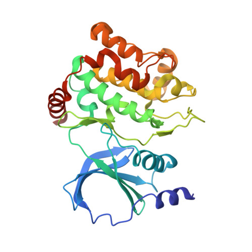
Serine/threonine-protein kinase PAK 1
A, B
300Homo sapiensMutation(s): 0 
Gene Names: PAK1
EC: 2.7.11.1
UniProt & NIH Common Fund Data Resources
Find proteins for Q13153 (Homo sapiens)
Explore Q13153 
Go to UniProtKB:  Q13153
PHAROS:  Q13153
GTEx:  ENSG00000149269 
Entity Groups  
Sequence Clusters30% Identity50% Identity70% Identity90% Identity95% Identity100% Identity
UniProt GroupQ13153
Sequence Annotations
Expand
  • Reference Sequence
Small Molecules
Ligands 1 Unique
IDChains Name / Formula / InChI Key2D Diagram3D Interactions
2OL
Query on 2OL

Download Ideal Coordinates CCD File 
C [auth A]1-({1-(2-aminopyrimidin-4-yl)-2-[(2-methoxyethyl)amino]-1H-benzimidazol-6-yl}ethynyl)cyclohexanol
C22 H26 N6 O2
RZXMIHOUHYSGJO-UHFFFAOYSA-N
Binding Affinity Annotations 
IDSourceBinding Affinity
2OL BindingDB:  4O0T Ki: 2900 (nM) from 1 assay(s)
IC50: min: 970, max: 1.00e+4 (nM) from 4 assay(s)
Experimental Data & Validation

Experimental Data

  • Method: X-RAY DIFFRACTION
  • Resolution: 2.60 Å
  • R-Value Free: 0.265 
  • R-Value Work: 0.214 
  • R-Value Observed: 0.217 
  • Space Group: P 1 21 1
Unit Cell:
Length ( Å )Angle ( ˚ )
a = 63.656α = 90
b = 79.17β = 106.97
c = 66.343γ = 90
Software Package:
Software NamePurpose
B3data collection
PHASERphasing
PHENIXrefinement
HKL-2000data reduction
SCALEPACKdata scaling

Structure Validation

View Full Validation Report


Ligand Structure Quality Assessment 


Entry History 

Deposition Data

Revision History  (Full details and data files)

  • Version 1.0: 2014-02-12
    Type: Initial release
  • Version 1.1: 2014-02-26
    Changes: Database references
  • Version 1.2: 2024-02-28
    Changes: Data collection, Database references, Derived calculations