4NQE

Crystal structure of TCR-MR1 ternary complex bound to 5-(2-oxoethylideneamino)-6-D-ribitylaminouracil


Experimental Data Snapshot

  • Method: X-RAY DIFFRACTION
  • Resolution: 2.10 Å
  • R-Value Free: 0.222 
  • R-Value Work: 0.182 
  • R-Value Observed: 0.184 

wwPDB Validation   3D Report Full Report


This is version 1.3 of the entry. See complete history


Literature

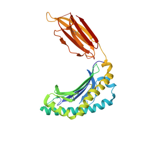
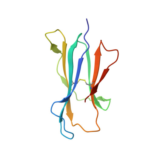
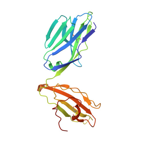
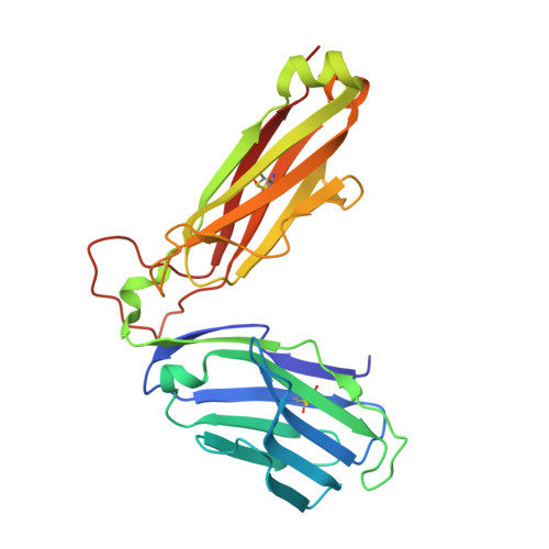
T-cell activation by transitory neo-antigens derived from distinct microbial pathways.

Corbett, A.J.Eckle, S.B.Birkinshaw, R.W.Liu, L.Patel, O.Mahony, J.Chen, Z.Reantragoon, R.Meehan, B.Cao, H.Williamson, N.A.Strugnell, R.A.Van Sinderen, D.Mak, J.Y.Fairlie, D.P.Kjer-Nielsen, L.Rossjohn, J.McCluskey, J.

(2014) Nature 509: 361-365

  • DOI: https://doi.org/10.1038/nature13160
  • Primary Citation of Related Structures:  
    4NQC, 4NQD, 4NQE

  • PubMed Abstract: 

    T cells discriminate between foreign and host molecules by recognizing distinct microbial molecules, predominantly peptides and lipids. Riboflavin precursors found in many bacteria and yeast also selectively activate mucosal-associated invariant T (MAIT) cells, an abundant population of innate-like T cells in humans. However, the genesis of these small organic molecules and their mode of presentation to MAIT cells by the major histocompatibility complex (MHC)-related protein MR1 (ref. 8) are not well understood. Here we show that MAIT-cell activation requires key genes encoding enzymes that form 5-amino-6-d-ribitylaminouracil (5-A-RU), an early intermediate in bacterial riboflavin synthesis. Although 5-A-RU does not bind MR1 or activate MAIT cells directly, it does form potent MAIT-activating antigens via non-enzymatic reactions with small molecules, such as glyoxal and methylglyoxal, which are derived from other metabolic pathways. The MAIT antigens formed by the reactions between 5-A-RU and glyoxal/methylglyoxal were simple adducts, 5-(2-oxoethylideneamino)-6-D-ribitylaminouracil (5-OE-RU) and 5-(2-oxopropylideneamino)-6-D-ribitylaminouracil (5-OP-RU), respectively, which bound to MR1 as shown by crystal structures of MAIT TCR ternary complexes. Although 5-OP-RU and 5-OE-RU are unstable intermediates, they became trapped by MR1 as reversible covalent Schiff base complexes. Mass spectra supported the capture by MR1 of 5-OP-RU and 5-OE-RU from bacterial cultures that activate MAIT cells, but not from non-activating bacteria, indicating that these MAIT antigens are present in a range of microbes. Thus, MR1 is able to capture, stabilize and present chemically unstable pyrimidine intermediates, which otherwise convert to lumazines, as potent antigens to MAIT cells. These pyrimidine adducts are microbial signatures for MAIT-cell immunosurveillance.


  • Organizational Affiliation

    1] Department of Microbiology and Immunology, Peter Doherty Institute for Infection and Immunity, University of Melbourne, Parkville, Victoria 3010, Australia [2].


Macromolecules
Find similar proteins by:  (by identity cutoff)  |  3D Structure
Entity ID: 1
MoleculeChains Sequence LengthOrganismDetailsImage
Major histocompatibility complex class I-related gene protein
A, C
271Homo sapiensMutation(s): 1 
Gene Names: MR1
UniProt & NIH Common Fund Data Resources
Find proteins for Q95460 (Homo sapiens)
Explore Q95460 
Go to UniProtKB:  Q95460
PHAROS:  Q95460
GTEx:  ENSG00000153029 
Entity Groups  
Sequence Clusters30% Identity50% Identity70% Identity90% Identity95% Identity100% Identity
UniProt GroupQ95460
Sequence Annotations
Expand
  • Reference Sequence
Find similar proteins by:  (by identity cutoff)  |  3D Structure
Entity ID: 2
MoleculeChains Sequence LengthOrganismDetailsImage
Beta-2-microglobulin
B, F
99Homo sapiensMutation(s): 0 
Gene Names: B2MCDABP0092HDCMA22P
UniProt & NIH Common Fund Data Resources
Find proteins for P61769 (Homo sapiens)
Explore P61769 
Go to UniProtKB:  P61769
PHAROS:  P61769
GTEx:  ENSG00000166710 
Entity Groups  
Sequence Clusters30% Identity50% Identity70% Identity90% Identity95% Identity100% Identity
UniProt GroupP61769
Sequence Annotations
Expand
  • Reference Sequence
Find similar proteins by:  (by identity cutoff)  |  3D Structure
Entity ID: 3
MoleculeChains Sequence LengthOrganismDetailsImage
TCR alpha
D, G
203Homo sapiensMutation(s): 0 
UniProt
Find proteins for Q6P4G7 (Homo sapiens)
Explore Q6P4G7 
Go to UniProtKB:  Q6P4G7
Entity Groups  
Sequence Clusters30% Identity50% Identity70% Identity90% Identity95% Identity100% Identity
UniProt GroupQ6P4G7
Sequence Annotations
Expand
  • Reference Sequence
Find similar proteins by:  (by identity cutoff)  |  3D Structure
Entity ID: 4
MoleculeChains Sequence LengthOrganismDetailsImage
TCR beta
E, H
245Homo sapiensMutation(s): 0 
UniProt
Find proteins for P01850 (Homo sapiens)
Explore P01850 
Go to UniProtKB:  P01850
Entity Groups  
Sequence Clusters30% Identity50% Identity70% Identity90% Identity95% Identity100% Identity
UniProt GroupP01850
Sequence Annotations
Expand
  • Reference Sequence
Small Molecules
Ligands 1 Unique
IDChains Name / Formula / InChI Key2D Diagram3D Interactions
2L4
Query on 2L4

Download Ideal Coordinates CCD File 
I [auth A],
J [auth C]
1-deoxy-1-({2,6-dioxo-5-[(E)-(2-oxoethylidene)amino]-1,2,3,6-tetrahydropyrimidin-4-yl}amino)-D-ribitol
C11 H16 N4 O7
PUEQUELBQOQOOV-GJQDMXJLSA-N
Experimental Data & Validation

Experimental Data

  • Method: X-RAY DIFFRACTION
  • Resolution: 2.10 Å
  • R-Value Free: 0.222 
  • R-Value Work: 0.182 
  • R-Value Observed: 0.184 
  • Space Group: C 1 2 1
Unit Cell:
Length ( Å )Angle ( ˚ )
a = 218.11α = 90
b = 70.6β = 104.63
c = 143.86γ = 90
Software Package:
Software NamePurpose
Blu-Icedata collection
PHASERphasing
BUSTERrefinement
MOSFLMdata reduction
SCALAdata scaling

Structure Validation

View Full Validation Report


Entry History 

Deposition Data

Revision History  (Full details and data files)

  • Version 1.0: 2014-04-16
    Type: Initial release
  • Version 1.1: 2014-05-28
    Changes: Database references
  • Version 1.2: 2014-11-19
    Changes: Structure summary
  • Version 1.3: 2020-02-19
    Changes: Advisory, Database references, Derived calculations