4MZV

Crystal structure of extracellular part of human EpCAM


Experimental Data Snapshot

  • Method: X-RAY DIFFRACTION
  • Resolution: 1.86 Å
  • R-Value Free: 0.249 
  • R-Value Work: 0.194 
  • R-Value Observed: 0.197 

wwPDB Validation   3D Report Full Report


Ligand Structure Quality Assessment 


This is version 2.0 of the entry. See complete history


Literature

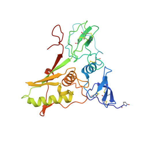
Crystal structure and its bearing towards an understanding of key biological functions of EpCAM.

Pavsic, M.Guncar, G.Djinovic-Carugo, K.Lenarcic, B.

(2014) Nat Commun 5: 4764-4764

  • DOI: https://doi.org/10.1038/ncomms5764
  • Primary Citation of Related Structures:  
    4MZV

  • PubMed Abstract: 

    EpCAM (epithelial cell adhesion molecule), a stem and carcinoma cell marker, is a cell surface protein involved in homotypic cell-cell adhesion via intercellular oligomerization and proliferative signalling via proteolytic cleavage. Despite its use as a diagnostic marker and being a drug target, structural details of this conserved vertebrate-exclusive protein remain unknown. Here we present the crystal structure of a heart-shaped dimer of the extracellular part of human EpCAM. The structure represents a cis-dimer that would form at cell surfaces and may provide the necessary structural foundation for the proposed EpCAM intercellular trans-tetramerization mediated by a membrane-distal region. By combining biochemical, biological and structural data on EpCAM, we show how proteolytic processing at various sites could influence structural integrity, oligomeric state and associated functionality of the molecule. We also describe the epitopes of this therapeutically important protein and explain the antigenicity of its regions.


  • Organizational Affiliation

    Department of Chemistry and Biochemistry, Faculty of Chemistry and Chemical Technology, University of Ljubljana, Večna pot 113, Ljubljana SI-1000, Slovenia.


Macromolecules
Find similar proteins by:  (by identity cutoff)  |  3D Structure
Entity ID: 1
MoleculeChains Sequence LengthOrganismDetailsImage
Epithelial cell adhesion molecule248Homo sapiensMutation(s): 3 
Gene Names: EPCAMGA733-2M1S2M4S1MIC18TACSTD1TROP1
UniProt & NIH Common Fund Data Resources
Find proteins for P16422 (Homo sapiens)
Explore P16422 
Go to UniProtKB:  P16422
PHAROS:  P16422
GTEx:  ENSG00000119888 
Entity Groups  
Sequence Clusters30% Identity50% Identity70% Identity90% Identity95% Identity100% Identity
UniProt GroupP16422
Sequence Annotations
Expand
  • Reference Sequence
Small Molecules
Ligands 1 Unique
IDChains Name / Formula / InChI Key2D Diagram3D Interactions
DMU
Query on DMU

Download Ideal Coordinates CCD File 
B [auth A]DECYL-BETA-D-MALTOPYRANOSIDE
C22 H42 O11
WOQQAWHSKSSAGF-WXFJLFHKSA-N
Modified Residues  1 Unique
IDChains TypeFormula2D DiagramParent
PCA
Query on PCA
A
L-PEPTIDE LINKINGC5 H7 N O3GLN
Experimental Data & Validation

Experimental Data

  • Method: X-RAY DIFFRACTION
  • Resolution: 1.86 Å
  • R-Value Free: 0.249 
  • R-Value Work: 0.194 
  • R-Value Observed: 0.197 
  • Space Group: C 1 2 1
Unit Cell:
Length ( Å )Angle ( ˚ )
a = 88.022α = 90
b = 50.385β = 128.33
c = 67.828γ = 90
Software Package:
Software NamePurpose
PROTEUM2data collection
SOLVEphasing
PHENIXrefinement
PROTEUM2data reduction
PROTEUM2data scaling

Structure Validation

View Full Validation Report


Ligand Structure Quality Assessment 


Entry History 

Deposition Data

Revision History  (Full details and data files)

  • Version 1.0: 2014-08-27
    Type: Initial release
  • Version 1.1: 2014-09-10
    Changes: Database references
  • Version 2.0: 2019-12-25
    Changes: Database references, Derived calculations, Polymer sequence