4MZQ

beta-Alanyl-CoA:Ammonia Lyase from Clostridium propionicum in complex with propionyl-CoA


Experimental Data Snapshot

  • Method: X-RAY DIFFRACTION
  • Resolution: 1.59 Å
  • R-Value Free: 0.213 
  • R-Value Work: 0.179 
  • R-Value Observed: 0.181 

wwPDB Validation   3D Report Full Report


Ligand Structure Quality Assessment 


This is version 1.3 of the entry. See complete history


Literature

High resolution crystal structure of Clostridium propionicum beta-alanyl-CoA:ammonia lyase, a new member of the "hot dog fold" protein superfamily.

Heine, A.Herrmann, G.Selmer, T.Terwesten, F.Buckel, W.Reuter, K.

(2014) Proteins 82: 2041-2053

  • DOI: https://doi.org/10.1002/prot.24557
  • Primary Citation of Related Structures:  
    4MTU, 4MZQ

  • PubMed Abstract: 

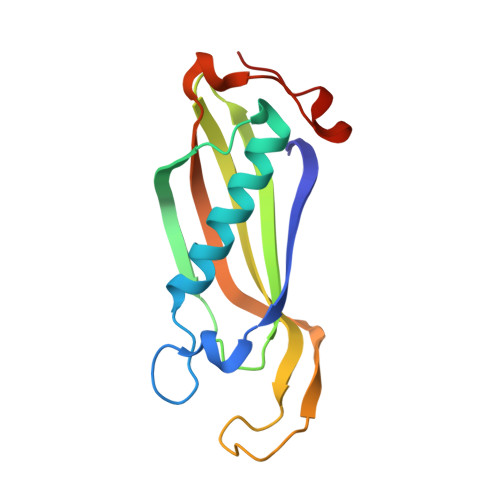
    Clostridium propionicum is the only organism known to ferment β-alanine, a constituent of coenzyme A (CoA) and the phosphopantetheinyl prosthetic group of holo-acyl carrier protein. The first step in the fermentation is a CoA-transfer to β-alanine. Subsequently, the resulting β-alanyl-CoA is deaminated by the enzyme β-alanyl-CoA:ammonia lyase (Acl) to reversibly form ammonia and acrylyl-CoA. We have determined the crystal structure of Acl in its apo-form at a resolution of 0.97 Å as well as in complex with CoA at a resolution of 1.59 Å. The structures reveal that the enyzme belongs to a superfamily of proteins exhibiting a so called "hot dog fold" which is characterized by a five-stranded antiparallel β-sheet with a long α-helix packed against it. The functional unit of all "hot dog fold" proteins is a homodimer containing two equivalent substrate binding sites which are established by the dimer interface. In the case of Acl, three functional dimers combine to a homohexamer strongly resembling the homohexamer formed by YciA-like acyl-CoA thioesterases. Here, we propose an enzymatic mechanism based on the crystal structure of the Acl·CoA complex and molecular docking.


  • Organizational Affiliation

    Institut für Pharmazeutische Chemie, Fachbereich Pharmazie, Philipps-Universität Marburg, Marbacher Weg 6, D-35032, Marburg, Germany.


Macromolecules
Find similar proteins by:  (by identity cutoff)  |  3D Structure
Entity ID: 1
MoleculeChains Sequence LengthOrganismDetailsImage
beta-Alanyl-CoA:Ammonia Lyase
A, B, C, D, E
A, B, C, D, E, F, G, H, I, J, K, L
144Anaerotignum propionicumMutation(s): 0 
UniProt
Find proteins for Q6KC22 (Anaerotignum propionicum)
Explore Q6KC22 
Go to UniProtKB:  Q6KC22
Entity Groups  
Sequence Clusters30% Identity50% Identity70% Identity90% Identity95% Identity100% Identity
UniProt GroupQ6KC22
Sequence Annotations
Expand
  • Reference Sequence
Small Molecules
Ligands 1 Unique
IDChains Name / Formula / InChI Key2D Diagram3D Interactions
1VU
Query on 1VU

Download Ideal Coordinates CCD File 
M [auth A]
N [auth B]
O [auth C]
P [auth D]
Q [auth E]
M [auth A],
N [auth B],
O [auth C],
P [auth D],
Q [auth E],
R [auth F],
S [auth G],
T [auth H],
U [auth I],
V [auth J],
W [auth K],
X [auth L]
propionyl Coenzyme A
C24 H40 N7 O17 P3 S
QAQREVBBADEHPA-IEXPHMLFSA-N
Experimental Data & Validation

Experimental Data

  • Method: X-RAY DIFFRACTION
  • Resolution: 1.59 Å
  • R-Value Free: 0.213 
  • R-Value Work: 0.179 
  • R-Value Observed: 0.181 
  • Space Group: P 1 21 1
Unit Cell:
Length ( Å )Angle ( ˚ )
a = 66.52α = 90
b = 110.965β = 89.98
c = 115.37γ = 90
Software Package:
Software NamePurpose
MAR345dtbdata collection
PHASERphasing
PHENIXrefinement
HKL-2000data reduction
HKL-2000data scaling

Structure Validation

View Full Validation Report


Ligand Structure Quality Assessment 


Entry History 

Deposition Data

Revision History  (Full details and data files)

  • Version 1.0: 2014-03-26
    Type: Initial release
  • Version 1.1: 2014-06-11
    Changes: Database references
  • Version 1.2: 2014-09-03
    Changes: Database references
  • Version 1.3: 2023-09-20
    Changes: Data collection, Database references, Derived calculations, Refinement description