4MY2

Crystal Structure of Norrin in fusion with Maltose Binding Protein


Experimental Data Snapshot

  • Method: X-RAY DIFFRACTION
  • Resolution: 2.40 Å
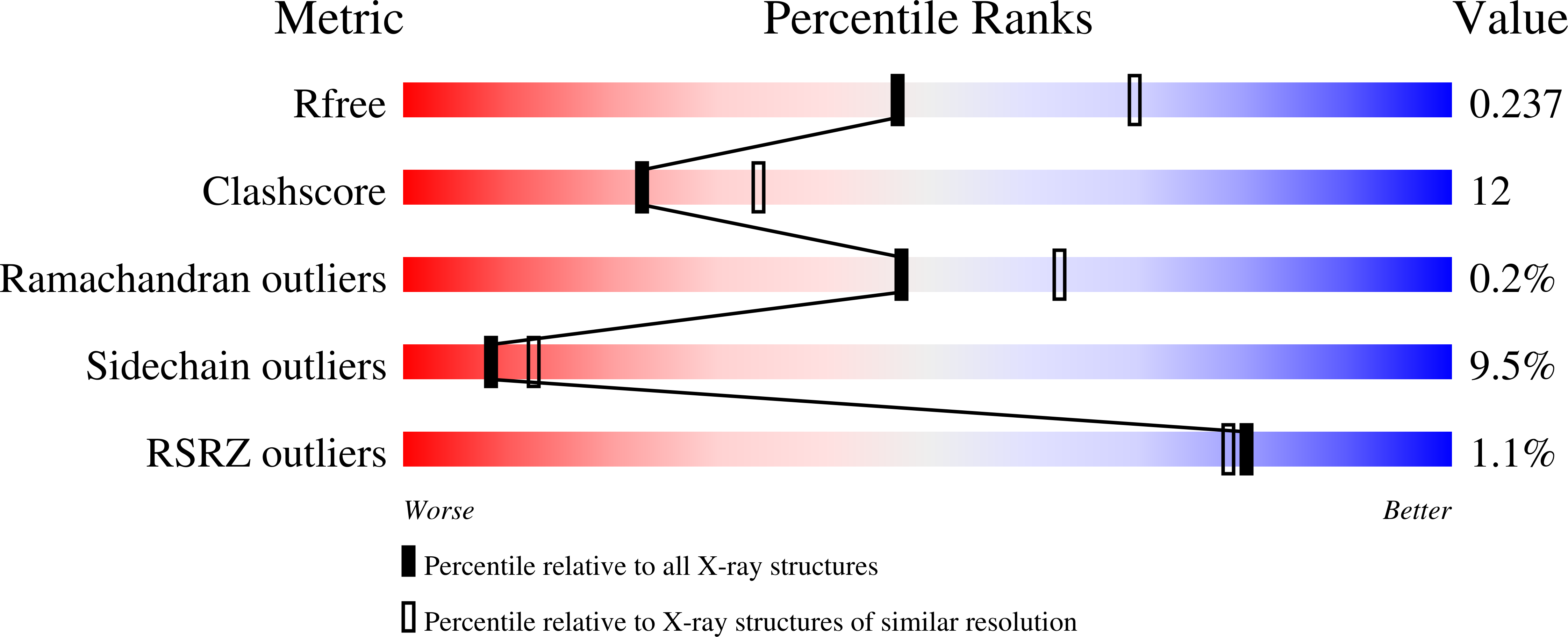
  • R-Value Free: 0.237 
  • R-Value Work: 0.207 
  • R-Value Observed: 0.208 

wwPDB Validation   3D Report Full Report


This is version 2.1 of the entry. See complete history


Literature

Structure and function of Norrin in assembly and activation of a Frizzled 4-Lrp5/6 complex.

Ke, J.Harikumar, K.G.Erice, C.Chen, C.Gu, X.Wang, L.Parker, N.Cheng, Z.Xu, W.Williams, B.O.Melcher, K.Miller, L.J.Xu, H.E.

(2013) Genes Dev 27: 2305-2319

  • DOI: https://doi.org/10.1101/gad.228544.113
  • Primary Citation of Related Structures:  
    4MY2

  • PubMed Abstract: 

    Norrin is a cysteine-rich growth factor that is required for angiogenesis in the eye, ear, brain, and female reproductive organs. It functions as an atypical Wnt ligand by specifically binding to the Frizzled 4 (Fz4) receptor. Here we report the crystal structure of Norrin, which reveals a unique dimeric structure with each monomer adopting a conserved cystine knot fold. Functional studies demonstrate that the novel Norrin dimer interface is required for Fz4 activation. Furthermore, we demonstrate that Norrin contains separate binding sites for Fz4 and for the Wnt ligand coreceptor Lrp5 (low-density lipoprotein-related protein 5) or Lrp6. Instead of inducing Fz4 dimerization, Norrin induces the formation of a ternary complex with Fz4 and Lrp5/6 by binding to their respective extracellular domains. These results provide crucial insights into the assembly and activation of the Norrin-Fz4-Lrp5/6 signaling complex.


  • Organizational Affiliation

    Laboratory of Structural Sciences, Van Andel Research Institute, Grand Rapids, Michigan 49503, USA;


Macromolecules
Find similar proteins by:  (by identity cutoff)  |  3D Structure
Entity ID: 1
MoleculeChains Sequence LengthOrganismDetailsImage
Maltose-binding periplasmic protein, Norrin fusion protein477Escherichia coli O157:H7Homo sapiens
This entity is chimeric
Mutation(s): 0 
Gene Names: malEZ5632ECs5017NDP
UniProt & NIH Common Fund Data Resources
Find proteins for P0AEX9 (Escherichia coli (strain K12))
Explore P0AEX9 
Go to UniProtKB:  P0AEX9
Find proteins for Q00604 (Homo sapiens)
Explore Q00604 
Go to UniProtKB:  Q00604
PHAROS:  Q00604
GTEx:  ENSG00000124479 
Entity Groups  
Sequence Clusters30% Identity50% Identity70% Identity90% Identity95% Identity100% Identity
UniProt GroupsP0AEX9Q00604
Sequence Annotations
Expand
  • Reference Sequence
Oligosaccharides

Help

Entity ID: 2
MoleculeChains Length2D Diagram Glycosylation3D Interactions
alpha-D-glucopyranose-(1-4)-alpha-D-glucopyranose
B
2N/A
Glycosylation Resources
GlyTouCan:  G07411ON
GlyCosmos:  G07411ON
Biologically Interesting Molecules (External Reference) 1 Unique
Experimental Data & Validation

Experimental Data

  • Method: X-RAY DIFFRACTION
  • Resolution: 2.40 Å
  • R-Value Free: 0.237 
  • R-Value Work: 0.207 
  • R-Value Observed: 0.208 
  • Space Group: P 21 21 2
Unit Cell:
Length ( Å )Angle ( ˚ )
a = 59.458α = 90
b = 79.022β = 90
c = 104.241γ = 90
Software Package:
Software NamePurpose
HKL-2000data collection
PHASERphasing
REFMACrefinement
HKL-2000data reduction
HKL-2000data scaling

Structure Validation

View Full Validation Report


Entry History 

Deposition Data

Revision History  (Full details and data files)

  • Version 1.0: 2013-11-13
    Type: Initial release
  • Version 1.1: 2013-11-20
    Changes: Database references
  • Version 1.2: 2017-08-09
    Changes: Refinement description, Source and taxonomy
  • Version 2.0: 2020-07-29
    Type: Remediation
    Reason: Carbohydrate remediation
    Changes: Advisory, Atomic model, Data collection, Database references, Derived calculations, Non-polymer description, Structure summary
  • Version 2.1: 2023-09-20
    Changes: Data collection, Database references, Refinement description, Structure summary