


Crystal Structure of Porcine C2 Domain of Blood Coagulation Factor VIII (FoldIt Target)
Spiegel, P.C., Brison, C.To be published.
Experimental Data Snapshot
Starting Model: experimental
View more details
wwPDB Validation 3D Report Full Report

Entity ID: 1 | |||||
|---|---|---|---|---|---|
| Molecule | Chains | Sequence Length | Organism | Details | Image |
| Coagulation factor VIII | A [auth M] | 177 | Sus scrofa | Mutation(s): 0 Gene Names: F8, CF8 Membrane Entity: Yes | 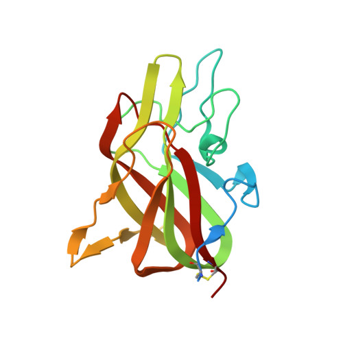 |
UniProt | |||||
Find proteins for P12263 (Sus scrofa) Explore P12263 Go to UniProtKB: P12263 | |||||
Entity Groups | |||||
| Sequence Clusters | 30% Identity50% Identity70% Identity90% Identity95% Identity100% Identity | ||||
| UniProt Group | P12263 | ||||
Sequence AnnotationsExpand | |||||
| |||||
| Ligands 1 Unique | |||||
|---|---|---|---|---|---|
| ID | Chains | Name / Formula / InChI Key | 2D Diagram | 3D Interactions | |
| GOL Query on GOL | B [auth M] | GLYCEROL C3 H8 O3 PEDCQBHIVMGVHV-UHFFFAOYSA-N |  | ||
| Length ( Å ) | Angle ( ˚ ) |
|---|---|
| a = 49.07 | α = 90 |
| b = 68.94 | β = 90 |
| c = 105.96 | γ = 90 |
| Software Name | Purpose |
|---|---|
| CrystalClear | data collection |
| Coot | model building |
| PHENIX | refinement |
| MOSFLM | data reduction |
| SCALA | data scaling |
RCSB PDB (citation) is hosted by
RCSB PDB is a member of the

