4MJ4

Human iduronidase apo structure P21 form


Experimental Data Snapshot

  • Method: X-RAY DIFFRACTION
  • Resolution: 2.17 Å
  • R-Value Free: 0.240 
  • R-Value Work: 0.201 
  • R-Value Observed: 0.203 

wwPDB Validation   3D Report Full Report


This is version 2.1 of the entry. See complete history


Literature

Insights into mucopolysaccharidosis I from the structure and action of alpha-L-iduronidase.

Bie, H.Yin, J.He, X.Kermode, A.R.Goddard-Borger, E.D.Withers, S.G.James, M.N.

(2013) Nat Chem Biol 9: 739-745

  • DOI: https://doi.org/10.1038/nchembio.1357
  • Primary Citation of Related Structures:  
    4KGJ, 4KGL, 4KH2, 4MJ2, 4MJ4

  • PubMed Abstract: 

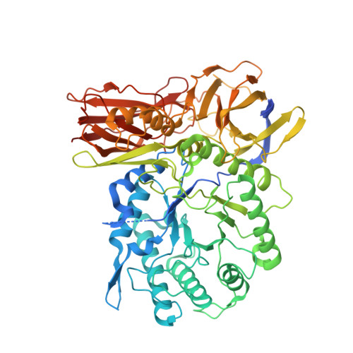
    Mucopolysaccharidosis type I (MPS I), caused by mutations in the gene encoding α-L-iduronidase (IDUA), is one of approximately 70 genetic disorders collectively known as the lysosomal storage diseases. To gain insight into the basis for MPS I, we crystallized human IDUA produced in an Arabidopsis thaliana cgl mutant. IDUA consists of a TIM barrel domain containing the catalytic site, a β-sandwich domain and a fibronectin-like domain. Structures of IDUA bound to iduronate analogs illustrate the Michaelis complex and reveal a (2,5)B conformation in the glycosyl-enzyme intermediate, which suggest a retaining double displacement reaction involving the nucleophilic Glu299 and the general acid/base Glu182. Unexpectedly, the N-glycan attached to Asn372 interacts with iduronate analogs in the active site and is required for enzymatic activity. Finally, these IDUA structures and biochemical analysis of the disease-relevant P533R mutation have enabled us to correlate the effects of mutations in IDUA to clinical phenotypes.


  • Organizational Affiliation

    1] Department of Biochemistry, University of Alberta, Edmonton, Alberta, Canada. [2] These authors contributed equally to this work.


Macromolecules
Find similar proteins by:  (by identity cutoff)  |  3D Structure
Entity ID: 1
MoleculeChains Sequence LengthOrganismDetailsImage
Alpha-L-iduronidase653Homo sapiensMutation(s): 0 
Gene Names: IDUA
EC: 3.2.1.76
UniProt & NIH Common Fund Data Resources
Find proteins for P35475 (Homo sapiens)
Explore P35475 
Go to UniProtKB:  P35475
PHAROS:  P35475
GTEx:  ENSG00000127415 
Entity Groups  
Sequence Clusters30% Identity50% Identity70% Identity90% Identity95% Identity100% Identity
UniProt GroupP35475
Sequence Annotations
Expand
  • Reference Sequence
Oligosaccharides

Help

Entity ID: 2
MoleculeChains Length2D Diagram Glycosylation3D Interactions
alpha-D-mannopyranose-(1-3)-[alpha-D-mannopyranose-(1-6)]beta-D-mannopyranose-(1-4)-2-acetamido-2-deoxy-beta-D-glucopyranose-(1-4)-2-acetamido-2-deoxy-beta-D-glucopyranose
B
5N-Glycosylation
Glycosylation Resources
GlyTouCan:  G22768VO
GlyCosmos:  G22768VO
GlyGen:  G22768VO
Experimental Data & Validation

Experimental Data

  • Method: X-RAY DIFFRACTION
  • Resolution: 2.17 Å
  • R-Value Free: 0.240 
  • R-Value Work: 0.201 
  • R-Value Observed: 0.203 
  • Space Group: P 1 21 1
Unit Cell:
Length ( Å )Angle ( ˚ )
a = 63.335α = 90
b = 70.507β = 92.1
c = 67.303γ = 90
Software Package:
Software NamePurpose
MxDCdata collection
PHASERphasing
REFMACrefinement
HKL-2000data reduction
SCALEPACKdata scaling

Structure Validation

View Full Validation Report


Entry History 

Deposition Data

Revision History  (Full details and data files)

  • Version 1.0: 2013-09-18
    Type: Initial release
  • Version 1.1: 2013-09-25
    Changes: Database references
  • Version 1.2: 2013-11-06
    Changes: Database references
  • Version 2.0: 2020-07-29
    Type: Remediation
    Reason: Carbohydrate remediation
    Changes: Atomic model, Data collection, Derived calculations, Structure summary
  • Version 2.1: 2023-09-20
    Changes: Data collection, Database references, Refinement description, Structure summary