


Crystal Structure of monomeric zebrafish beta-2-microglobulin
Wu, P.H., Lai, M.H., Li, C.H., Chang, C.C., Chen, C.J.To be published.
Experimental Data Snapshot
wwPDB Validation 3D Report Full Report

Entity ID: 1 | |||||
|---|---|---|---|---|---|
| Molecule | Chains | Sequence Length | Organism | Details | Image |
| Beta-2-microglobulin | 107 | Danio rerio | Mutation(s): 0 Gene Names: b2m, beta-2-microglobulin | 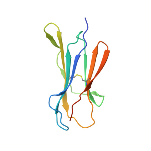 | |
UniProt | |||||
Find proteins for Q04475 (Danio rerio) Explore Q04475 Go to UniProtKB: Q04475 | |||||
Entity Groups | |||||
| Sequence Clusters | 30% Identity50% Identity70% Identity90% Identity95% Identity100% Identity | ||||
| UniProt Group | Q04475 | ||||
Sequence AnnotationsExpand | |||||
| |||||
| Length ( Å ) | Angle ( ˚ ) |
|---|---|
| a = 38.084 | α = 90 |
| b = 50.926 | β = 90 |
| c = 54.07 | γ = 90 |
| Software Name | Purpose |
|---|---|
| HKL-2000 | data collection |
| AMoRE | phasing |
| REFMAC | refinement |
| HKL-2000 | data reduction |
| HKL-2000 | data scaling |
RCSB PDB (citation) is hosted by
RCSB PDB is a member of the

