4M1U

The crystal structure of Stx2 and a disaccharide ligand


Experimental Data Snapshot

  • Method: X-RAY DIFFRACTION
  • Resolution: 1.56 Å
  • R-Value Free: 0.192 
  • R-Value Work: 0.170 
  • R-Value Observed: 0.171 

wwPDB Validation   3D Report Full Report


Ligand Structure Quality Assessment 


This is version 2.1 of the entry. See complete history


Literature

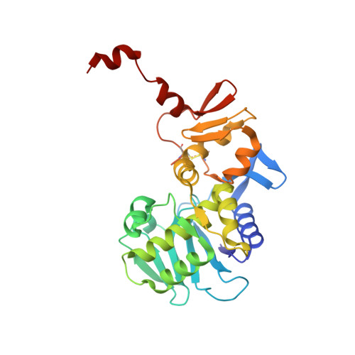
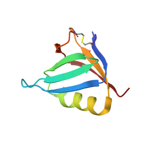
The crystal structure of shiga toxin type 2 with bound disaccharide guides the design of a heterobifunctional toxin inhibitor.

Jacobson, J.M.Yin, J.Kitov, P.I.Mulvey, G.Griener, T.P.James, M.N.Armstrong, G.Bundle, D.R.

(2014) J Biol Chem 289: 885-894

  • DOI: https://doi.org/10.1074/jbc.M113.518886
  • Primary Citation of Related Structures:  
    4M1U

  • PubMed Abstract: 

    Shiga toxin type 2 (Stx2a) is clinically most closely associated with enterohemorrhagic E. coli O157:H7-mediated hemorrhagic colitis that sometimes progresses to hemolytic-uremic syndrome. The ability to express the toxin has been acquired by other Escherichia coli strains, and outbreaks of food poisoning have caused significant mortality rates as, for example, in the 2011 outbreak in northern Germany. Stx2a, an AB5 toxin, gains entry into human cells via the glycosphingolipid receptor Gb3. We have determined the first crystal structure of a disaccharide analog of Gb3 bound to the B5 pentamer of Stx2a holotoxin. In this Gb3 analog,-GalNAc replaces the terminal-Gal residue. This co-crystal structure confirms previous inferences that two of the primary binding sites identified in theB5 pentamer of Stx1 are also functional in Stx2a. This knowledge provides a rationale for the synthesis and evaluation of heterobifunctional antagonists for E. coli toxins that target Stx2a. Incorporation of GalNAc Gb3 trisaccharide in a heterobifunctional ligand with an attached pyruvate acetal, a ligand for human amyloid P component, and conjugation to poly[acrylamide-co-(3-azidopropylmethacrylamide)] produced a polymer that neutralized Stx2a in a mouse model of Shigatoxemia.


  • Organizational Affiliation

    From the Department of Chemistry, Alberta Glycomics Centre, University of Alberta, Edmonton, Alberta T6G 2G2, Canada.


Macromolecules
Find similar proteins by:  (by identity cutoff)  |  3D Structure
Entity ID: 1
MoleculeChains Sequence LengthOrganismDetailsImage
Shiga toxin 2 A-subunit297Escherichia coli O157:H7Mutation(s): 0 
Gene Names: ECs1205stx 2 A-subunitstx2 A-subunitstx2AZ1464
EC: 3.2.2.22
UniProt
Find proteins for Q7DI68 (Escherichia coli O157:H7)
Explore Q7DI68 
Go to UniProtKB:  Q7DI68
Entity Groups  
Sequence Clusters30% Identity50% Identity70% Identity90% Identity95% Identity100% Identity
UniProt GroupQ7DI68
Sequence Annotations
Expand
  • Reference Sequence
Find similar proteins by:  (by identity cutoff)  |  3D Structure
Entity ID: 2
MoleculeChains Sequence LengthOrganismDetailsImage
Shiga toxin 2 B subunit
B, C, D, E, F
70Escherichia coliMutation(s): 0 
Gene Names: stx2Bstx2dBstx2vBstxB2stxIIvtx2B
UniProt
Find proteins for Q7DJJ2 (Escherichia coli)
Explore Q7DJJ2 
Go to UniProtKB:  Q7DJJ2
Entity Groups  
Sequence Clusters30% Identity50% Identity70% Identity90% Identity95% Identity100% Identity
UniProt GroupQ7DJJ2
Sequence Annotations
Expand
  • Reference Sequence
Oligosaccharides

Help

Entity ID: 3
MoleculeChains Length2D Diagram Glycosylation3D Interactions
2-acetamido-2-deoxy-alpha-D-galactopyranose-(1-4)-methyl beta-D-galactopyranoside
G, H
2N/A
Glycosylation Resources
GlyTouCan:  G78497LG
GlyCosmos:  G78497LG
Experimental Data & Validation

Experimental Data

  • Method: X-RAY DIFFRACTION
  • Resolution: 1.56 Å
  • R-Value Free: 0.192 
  • R-Value Work: 0.170 
  • R-Value Observed: 0.171 
  • Space Group: P 61
Unit Cell:
Length ( Å )Angle ( ˚ )
a = 146.25α = 90
b = 146.25β = 90
c = 60.31γ = 120
Software Package:
Software NamePurpose
MxDCdata collection
PHASERphasing
REFMACrefinement
XDSdata reduction
XDSdata scaling

Structure Validation

View Full Validation Report


Ligand Structure Quality Assessment 


Entry History 

Deposition Data

Revision History  (Full details and data files)

  • Version 1.0: 2013-11-20
    Type: Initial release
  • Version 1.1: 2013-12-04
    Changes: Database references
  • Version 1.2: 2014-02-19
    Changes: Database references
  • Version 2.0: 2020-07-29
    Type: Remediation
    Reason: Carbohydrate remediation
    Changes: Atomic model, Data collection, Derived calculations, Structure summary
  • Version 2.1: 2023-09-20
    Changes: Data collection, Database references, Refinement description, Structure summary