Structural Basis for Assembly of the Mn(IV)/Fe(III) Cofactor in the Class Ic Ribonucleotide Reductase from Chlamydia trachomatis.
Dassama, L.M., Krebs, C., Bollinger, J.M., Rosenzweig, A.C., Boal, A.K.(2013) Biochemistry 52: 6424-6436
- PubMed: 23924396
- DOI: https://doi.org/10.1021/bi400819x
- Primary Citation of Related Structures:
4M1H, 4M1I - PubMed Abstract:
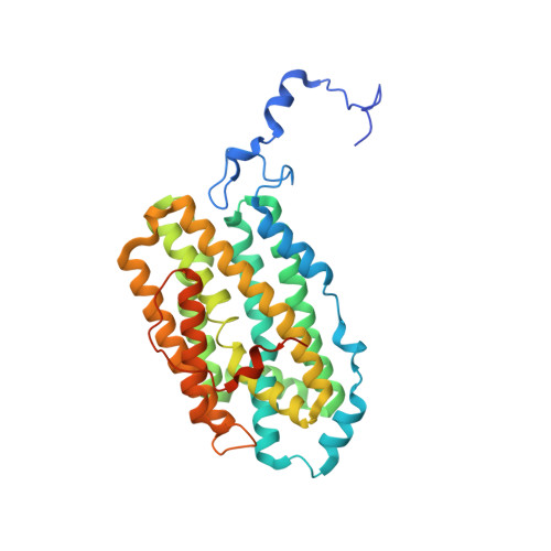
The class Ic ribonucleotide reductase (RNR) from Chlamydia trachomatis (Ct) employs a Mn(IV)/Fe(III) cofactor in each monomer of its β2 subunit to initiate nucleotide reduction. The cofactor forms by reaction of Mn(II)/Fe(II)-β2 with O2. Previously, in vitro cofactor assembly from apo β2 and divalent metal ions produced a mixture of two forms, with Mn at site 1 (Mn(IV)/Fe(III)) or site 2 (Fe(III)/Mn(IV)), of which the more active Mn(IV)/Fe(III) product predominates. Here we have addressed the basis for metal site selectivity by determining X-ray crystal structures of apo, Mn(II), and Mn(II)/Fe(II) complexes of Ct β2. A structure obtained anaerobically with equimolar Mn(II), Fe(II), and apoprotein reveals exclusive incorporation of Mn(II) at site 1 and Fe(II) at site 2, in contrast to the more modest site selectivity achieved previously. Site specificity is controlled thermodynamically by the apoprotein structure, as only minor adjustments of ligands occur upon metal binding. Additional structures imply that, by itself, Mn(II) binds in either site. Together, the structures are consistent with a model for in vitro cofactor assembly in which Fe(II) specificity for site 2 drives assembly of the appropriately configured heterobimetallic center, provided that Fe(II) is substoichiometric. This model suggests that use of a Mn(IV)/Fe(III) cofactor in vivo could be an adaptation to Fe(II) limitation. A 1.8 Å resolution model of the Mn(II)/Fe(II)-β2 complex reveals additional structural determinants for activation of the cofactor, including a proposed site for side-on (η(2)) addition of O2 to Fe(II) and a short (3.2 Å) Mn(II)-Fe(II) interionic distance, promoting formation of the Mn(IV)/Fe(IV) activation intermediate.
Organizational Affiliation:
Department of Chemistry and Department of Biochemistry and Molecular Biology, The Pennsylvania State University , University Park, Pennsylvania 16802, United States.