4LOO

Structural basis of autoactivation of p38 alpha induced by TAB1 (Monoclinic crystal form)


Experimental Data Snapshot

  • Method: X-RAY DIFFRACTION
  • Resolution: 1.95 Å
  • R-Value Free: 0.250 
  • R-Value Work: 0.198 
  • R-Value Observed: 0.200 

wwPDB Validation   3D Report Full Report


Ligand Structure Quality Assessment 


This is version 1.4 of the entry. See complete history


Literature

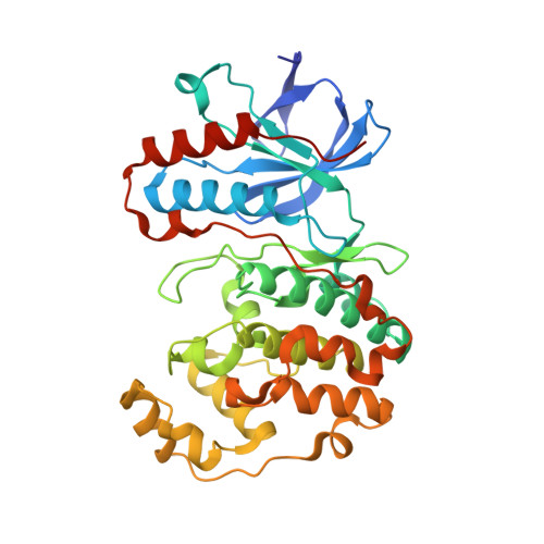
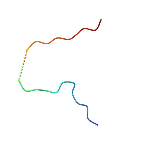
Mechanism and consequence of the autoactivation of p38 alpha mitogen-activated protein kinase promoted by TAB1.

De Nicola, G.F.Martin, E.D.Chaikuad, A.Bassi, R.Clark, J.Martino, L.Verma, S.Sicard, P.Tata, R.Atkinson, R.A.Knapp, S.Conte, M.R.Marber, M.S.

(2013) Nat Struct Mol Biol 20: 1182-1190

  • DOI: https://doi.org/10.1038/nsmb.2668
  • Primary Citation of Related Structures:  
    4LOO, 4LOP, 4LOQ

  • PubMed Abstract: 

    p38α mitogen-activated protein kinase (p38α) is activated by a variety of mechanisms, including autophosphorylation initiated by TGFβ-activated kinase 1 binding protein 1 (TAB1) during myocardial ischemia and other stresses. Chemical-genetic approaches and coexpression in mammalian, bacterial and cell-free systems revealed that mouse p38α autophosphorylation occurs in cis by direct interaction with TAB1(371-416). In isolated rat cardiac myocytes and perfused mouse hearts, TAT-TAB1(371-416) rapidly activates p38 and profoundly perturbs function. Crystal structures and characterization in solution revealed a bipartite docking site for TAB1 in the p38α C-terminal kinase lobe. TAB1 binding stabilizes active p38α and induces rearrangements within the activation segment by helical extension of the Thr-Gly-Tyr motif, allowing autophosphorylation in cis. Interference with p38α recognition by TAB1 abolishes its cardiac toxicity. Such intervention could potentially circumvent the drawbacks of clinical pharmacological inhibitors of p38 catalytic activity.


  • Organizational Affiliation

    King's College London British Heart Foundation Centre of Excellence. The Rayne Institute, St Thomas' Hospital Campus, London, SE1 7EH, UK.


Macromolecules
Find similar proteins by:  (by identity cutoff)  |  3D Structure
Entity ID: 1
MoleculeChains Sequence LengthOrganismDetailsImage
Mitogen-activated protein kinase 14361Mus musculusMutation(s): 0 
Gene Names: MAPK14Crk1Csbp1Csbp2
EC: 2.7.11.24
UniProt
Find proteins for P47811 (Mus musculus)
Explore P47811 
Go to UniProtKB:  P47811
Entity Groups  
Sequence Clusters30% Identity50% Identity70% Identity90% Identity95% Identity100% Identity
UniProt GroupP47811
Sequence Annotations
Expand
  • Reference Sequence
Find similar proteins by:  (by identity cutoff)  |  3D Structure
Entity ID: 2
MoleculeChains Sequence LengthOrganismDetailsImage
TGF-beta-activated kinase 1 and MAP3K7-binding protein 129Mus musculusMutation(s): 0 
UniProt
Find proteins for Q8CF89 (Mus musculus)
Explore Q8CF89 
Go to UniProtKB:  Q8CF89
Entity Groups  
Sequence Clusters30% Identity50% Identity70% Identity90% Identity95% Identity100% Identity
UniProt GroupQ8CF89
Sequence Annotations
Expand
  • Reference Sequence
Small Molecules
Ligands 1 Unique
IDChains Name / Formula / InChI Key2D Diagram3D Interactions
SB4
Query on SB4

Download Ideal Coordinates CCD File 
C [auth A]4-(4-FLUOROPHENYL)-1-(4-PIPERIDINYL)-5-(2-AMINO-4-PYRIMIDINYL)-IMIDAZOLE
C18 H19 F N6
VSPFURGQAYMVAN-UHFFFAOYSA-N
Binding Affinity Annotations 
IDSourceBinding Affinity
SB4 PDBBind:  4LOO Kd: 7 (nM) from 1 assay(s)
BindingDB:  4LOO IC50: min: 19, max: 20 (nM) from 2 assay(s)
Experimental Data & Validation

Experimental Data

  • Method: X-RAY DIFFRACTION
  • Resolution: 1.95 Å
  • R-Value Free: 0.250 
  • R-Value Work: 0.198 
  • R-Value Observed: 0.200 
  • Space Group: P 1 21 1
Unit Cell:
Length ( Å )Angle ( ˚ )
a = 43.34α = 90
b = 73.58β = 91.15
c = 59.19γ = 90
Software Package:
Software NamePurpose
GDAdata collection
PHASERphasing
REFMACrefinement
MOSFLMdata reduction
SCALAdata scaling

Structure Validation

View Full Validation Report


Ligand Structure Quality Assessment 


Entry History 

Revision History  (Full details and data files)

  • Version 1.0: 2013-08-21
    Type: Initial release
  • Version 1.1: 2013-09-11
    Changes: Database references
  • Version 1.2: 2013-10-02
    Changes: Database references
  • Version 1.3: 2013-10-23
    Changes: Database references
  • Version 1.4: 2023-09-20
    Changes: Data collection, Database references, Derived calculations, Refinement description