4LHQ

Ricin A chain bound to camelid nanobody (VHH8)


Experimental Data Snapshot

  • Method: X-RAY DIFFRACTION
  • Resolution: 2.30 Å
  • R-Value Free: 0.248 
  • R-Value Work: 0.192 
  • R-Value Observed: 0.193 

wwPDB Validation   3D Report Full Report


This is version 1.6 of the entry. See complete history


Literature

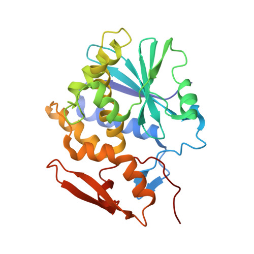
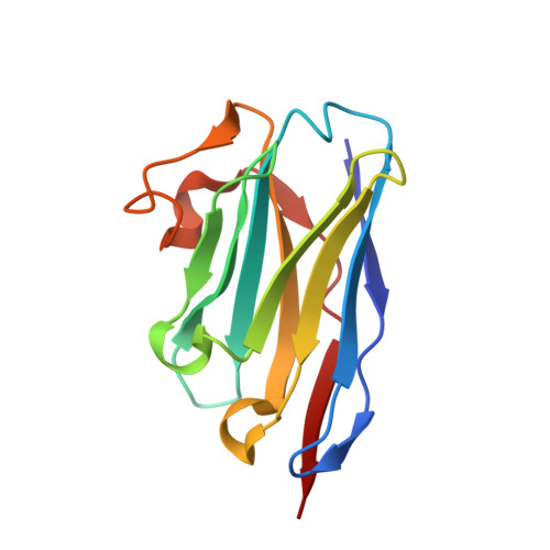
Crystal Structures of Ricin Toxin's Enzymatic Subunit (RTA) in Complex with Neutralizing and Non-Neutralizing Single-Chain Antibodies.

Rudolph, M.J.Vance, D.J.Cheung, J.Franklin, M.C.Burshteyn, F.Cassidy, M.S.Gary, E.N.Herrera, C.Shoemaker, C.B.Mantis, N.J.

(2014) J Mol Biol 426: 3057-3068

  • DOI: https://doi.org/10.1016/j.jmb.2014.05.026
  • Primary Citation of Related Structures:  
    4LGP, 4LGR, 4LGS, 4LHJ, 4LHQ

  • PubMed Abstract: 

    Ricin is a select agent toxin and a member of the RNA N-glycosidase family of medically important plant and bacterial ribosome-inactivating proteins. In this study, we determined X-ray crystal structures of the enzymatic subunit of ricin (RTA) in complex with the antigen binding domains (VHH) of five unique single-chain monoclonal antibodies that differ in their respective toxin-neutralizing activities. None of the VHHs made direct contact with residues involved in RTA's RNA N-glycosidase activity or induced notable allosteric changes in the toxin's subunit. Rather, the five VHHs had overlapping structural epitopes on the surface of the toxin and differed in the degree to which they made contact with prominent structural elements in two folding domains of the RTA. In general, RTA interactions were influenced most by the VHH CDR3 (CDR, complementarity-determining region) elements, with the most potent neutralizing antibody having the shortest and most conformationally constrained CDR3. These structures provide unique insights into the mechanisms underlying toxin neutralization and provide critically important information required for the rational design of ricin toxin subunit vaccines.


  • Organizational Affiliation

    New York Structural Biology Center, New York, NY 10027, USA. Electronic address: mrudolph@nysbc.org.


Macromolecules
Find similar proteins by:  (by identity cutoff)  |  3D Structure
Entity ID: 1
MoleculeChains Sequence LengthOrganismDetailsImage
Ricin
A, C
260Ricinus communisMutation(s): 0 
EC: 3.2.2.22
UniProt
Find proteins for P02879 (Ricinus communis)
Explore P02879 
Go to UniProtKB:  P02879
Entity Groups  
Sequence Clusters30% Identity50% Identity70% Identity90% Identity95% Identity100% Identity
UniProt GroupP02879
Sequence Annotations
Expand
  • Reference Sequence
Find similar proteins by:  (by identity cutoff)  |  3D Structure
Entity ID: 2
MoleculeChains Sequence LengthOrganismDetailsImage
Camelid nanobody
B, D
128Vicugna pacosMutation(s): 0 
Entity Groups  
Sequence Clusters30% Identity50% Identity70% Identity90% Identity95% Identity100% Identity
Sequence Annotations
Expand
  • Reference Sequence
Experimental Data & Validation

Experimental Data

  • Method: X-RAY DIFFRACTION
  • Resolution: 2.30 Å
  • R-Value Free: 0.248 
  • R-Value Work: 0.192 
  • R-Value Observed: 0.193 
  • Space Group: P 41
Unit Cell:
Length ( Å )Angle ( ˚ )
a = 49.048α = 90
b = 49.048β = 90
c = 339.595γ = 90
Software Package:
Software NamePurpose
SCALEPACKdata scaling
PHASERphasing
PHENIXrefinement
PDB_EXTRACTdata extraction
HKL-2000data collection
DENZOdata reduction

Structure Validation

View Full Validation Report


Entry History 

Deposition Data

Revision History  (Full details and data files)

  • Version 1.0: 2014-06-11
    Type: Initial release
  • Version 1.1: 2014-07-02
    Changes: Database references
  • Version 1.2: 2014-07-30
    Changes: Database references
  • Version 1.3: 2014-08-20
    Changes: Database references
  • Version 1.4: 2017-03-15
    Changes: Source and taxonomy
  • Version 1.5: 2017-11-15
    Changes: Refinement description
  • Version 1.6: 2024-02-28
    Changes: Data collection, Database references