4LF1

Hexameric Form II RuBisCO from Rhodopseudomonas palustris, activated and complexed with 2-CABP


Experimental Data Snapshot

  • Method: X-RAY DIFFRACTION
  • Resolution: 2.38 Å
  • R-Value Free: 0.315 
  • R-Value Work: 0.263 
  • R-Value Observed: 0.266 

wwPDB Validation   3D Report Full Report


Ligand Structure Quality Assessment 


This is version 1.1 of the entry. See complete history


Literature

Structure-function studies with the unique hexameric form II ribulose-1,5-bisphosphate carboxylase/oxygenase (Rubisco) from Rhodopseudomonas palustris.

Satagopan, S.Chan, S.Perry, L.J.Tabita, F.R.

(2014) J Biol Chem 289: 21433-21450

  • DOI: https://doi.org/10.1074/jbc.M114.578625
  • Primary Citation of Related Structures:  
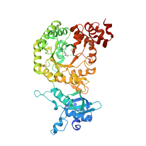
    4LF1, 4LF2

  • PubMed Abstract: 

    The first x-ray crystal structure has been solved for an activated transition-state analog-bound form II ribulose-1,5-bisphosphate carboxylase/oxygenase (Rubisco). This enzyme, from Rhodopseudomonas palustris, assembles as a unique hexamer with three pairs of catalytic large subunit homodimers around a central 3-fold symmetry axis. This oligomer arrangement is unique among all known Rubisco structures, including the form II homolog from Rhodospirillum rubrum. The presence of a transition-state analog in the active site locked the activated enzyme in a "closed" conformation and revealed the positions of critical active site residues during catalysis. Functional roles of two form II-specific residues (Ile(165) and Met(331)) near the active site were examined via site-directed mutagenesis. Substitutions at these residues affect function but not the ability of the enzyme to assemble. Random mutagenesis and suppressor selection in a Rubisco deletion strain of Rhodobacter capsulatus identified a residue in the amino terminus of one subunit (Ala(47)) that compensated for a negative change near the active site of a neighboring subunit. In addition, substitution of the native carboxyl-terminal sequence with the last few dissimilar residues from the related R. rubrum homolog increased the enzyme's kcat for carboxylation. However, replacement of a longer carboxyl-terminal sequence with termini from either a form III or a form I enzyme, which varied both in length and sequence, resulted in complete loss of function. From these studies, it is evident that a number of subtle interactions near the active site and the carboxyl terminus account for functional differences between the different forms of Rubiscos found in nature.


  • Organizational Affiliation

    From the Department of Microbiology, The Ohio State University, Columbus, Ohio 43210-1292 and.


Macromolecules
Find similar proteins by:  (by identity cutoff)  |  3D Structure
Entity ID: 1
MoleculeChains Sequence LengthOrganismDetailsImage
Ribulose bisphosphate carboxylase
A, B, C, D, E
A, B, C, D, E, F
481Rhodopseudomonas palustris CGA009Mutation(s): 0 
Gene Names: cbbMRPA4641Rpal_5122
EC: 4.1.1.39
UniProt
Find proteins for Q6N0W9 (Rhodopseudomonas palustris (strain ATCC BAA-98 / CGA009))
Explore Q6N0W9 
Go to UniProtKB:  Q6N0W9
Entity Groups  
Sequence Clusters30% Identity50% Identity70% Identity90% Identity95% Identity100% Identity
UniProt GroupQ6N0W9
Sequence Annotations
Expand
  • Reference Sequence
Small Molecules
Ligands 2 Unique
IDChains Name / Formula / InChI Key2D Diagram3D Interactions
CAP
Query on CAP

Download Ideal Coordinates CCD File 
G [auth A]
I [auth B]
K [auth C]
M [auth D]
O [auth E]
G [auth A],
I [auth B],
K [auth C],
M [auth D],
O [auth E],
Q [auth F]
2-CARBOXYARABINITOL-1,5-DIPHOSPHATE
C6 H14 O13 P2
ITHCSGCUQDMYAI-ZMIZWQJLSA-N
MG
Query on MG

Download Ideal Coordinates CCD File 
H [auth A]
J [auth B]
L [auth C]
N [auth D]
P [auth E]
H [auth A],
J [auth B],
L [auth C],
N [auth D],
P [auth E],
R [auth F]
MAGNESIUM ION
Mg
JLVVSXFLKOJNIY-UHFFFAOYSA-N
Modified Residues  1 Unique
IDChains TypeFormula2D DiagramParent
KCX
Query on KCX
A, B, C, D, E
A, B, C, D, E, F
L-PEPTIDE LINKINGC7 H14 N2 O4LYS
Experimental Data & Validation

Experimental Data

  • Method: X-RAY DIFFRACTION
  • Resolution: 2.38 Å
  • R-Value Free: 0.315 
  • R-Value Work: 0.263 
  • R-Value Observed: 0.266 
  • Space Group: P 1
Unit Cell:
Length ( Å )Angle ( ˚ )
a = 75.033α = 66.51
b = 100.72β = 108.32
c = 100.69γ = 95.41
Software Package:
Software NamePurpose
DENZOdata reduction
SCALEPACKdata scaling
EPMRphasing
REFMACrefinement
PDB_EXTRACTdata extraction
HKL-2000data collection

Structure Validation

View Full Validation Report


Ligand Structure Quality Assessment 


Entry History 

Deposition Data

Revision History  (Full details and data files)

  • Version 1.0: 2014-06-25
    Type: Initial release
  • Version 1.1: 2016-07-20
    Changes: Database references