Structural basis of the substrate specificity of cytidine deaminase superfamily Guanine deaminase
Bitra, A., Biswas, A., Anand, R.(2013) Biochemistry 52: 8106-8114
- PubMed: 24083949
- DOI: https://doi.org/10.1021/bi400818e
- Primary Citation of Related Structures:
4LC5, 4LCN, 4LCO, 4LCP, 4LD2, 4LD4 - PubMed Abstract:
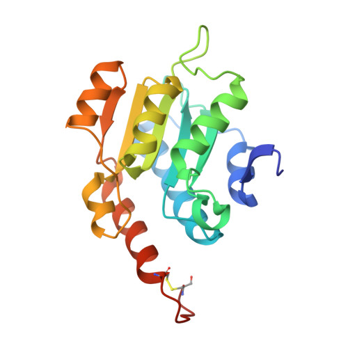
Guanine deaminases (GDs) are important enzymes involved in purine metabolism as well as nucleotide anabolism pathways that exhibit a high degree of fidelity. Here, the structural basis of the substrate specificity of GDs was investigated by determining a series of X-ray structures of NE0047 (GD from Nitrosomonas europaea) with nucleobase analogues and nucleosides. The structures demonstrated that the interactions in the GD active site are tailor-made to accommodate only guanine and any substitutions in the purine ring or introduction of a pyrimidine ring results in rearrangement of the bases in a catalytically unfavorable orientation, away from the proton shuttling residue E143. In addition, X-ray structural studies performed on cytidine revealed that although it binds in an optimal conformation, its deamination does not occur because of the inability of the enzyme to orchestrate the closure of the catalytically important C-terminal loop (residues 181-189). Isothermal calorimetry measurements established that these nucleoside moieties also disrupt the sequential mode of ligand binding, thereby abrogating all intersubunit communication. Intriguingly, it was recently discovered that GDs can also serve as endogenous ammeline deaminases, although it is structurally nonhomologous with guanine. To understand the mechanism of dual-substrate specificity, the structure of NE0047 in complex with ammeline was determined to a resolution of 2.7 Å. The structure revealed that ammeline not only fits in the active site in a catalytically favorable orientation but also allows for closure of the C-terminal loop.
Organizational Affiliation:
Department of Chemistry, Indian Institute of Technology , Mumbai 400076, India.