Converting NAD-Specific Inositol Dehydrogenase to an Efficient NADP-Selective Catalyst, with a Surprising Twist.
Zheng, H., Bertwistle, D., Sanders, D.A., Palmer, D.R.(2013) Biochemistry 52: 5876-5883
- PubMed: 23952058
- DOI: https://doi.org/10.1021/bi400821s
- Primary Citation of Related Structures:
4L8V, 4L9R - PubMed Abstract:
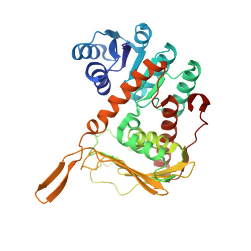
myo-Inositol dehydrogenase (IDH, EC 1.1.1.18) from Bacillus subtilis converts myo-inositol to scyllo-inosose and is strictly dependent on NAD for activity. We sought to alter the coenzyme specificity to generate an NADP-dependent enzyme in order to enhance our understanding of coenzyme selectivity and to create an enzyme capable of recycling NADP in biocatalytic processes. Examination of available structural information related to the GFO/MocA/IDH family of dehydrogenases and precedents for altering coenzyme selectivity allowed us to select residues for substitution, and nine single, double, and triple mutants were constructed. Mutagenesis experiments with B. subtilis IDH proved extremely successful; the double mutant D35S/V36R preferred NADP to NAD by a factor of 5. This mutant is an excellent catalyst with a second-order rate constant with respect to NADP of 370 000 s⁻¹ M⁻¹, and the triple mutant A12K/D35S/V36R had a value of 570 000 s⁻¹ M⁻¹, higher than that of the wild-type IDH with NAD. The high-resolution X-ray crystal structure of the double mutant A12K/D35S was solved in complex with NADP. Surprisingly, the binding of the coenzyme is altered such that although the nicotinamide ring maintains the required position for catalysis, the coenzyme has twisted by nearly 90°, so the adenine moiety no longer binds to a hydrophobic cleft in the Rossmann fold as in the wild-type enzyme. This change in binding conformation has not previously been observed in mutated dehydrogenases.
Organizational Affiliation:
Department of Chemistry, University of Saskatchewan, 110 Science Place, Saskatoon, Saskatchewan S7N 5C9, Canada.