4KW1

Structure of a/egypt/n03072/2010 h5 ha


Experimental Data Snapshot

  • Method: X-RAY DIFFRACTION
  • Resolution: 2.50 Å
  • R-Value Free: 0.227 
  • R-Value Work: 0.193 
  • R-Value Observed: 0.194 

wwPDB Validation   3D Report Full Report


Ligand Structure Quality Assessment 


This is version 2.0 of the entry. See complete history


Literature

Structural and antigenic variation among diverse clade 2 H5N1 viruses.

Shore, D.A.Yang, H.Balish, A.L.Shepard, S.S.Carney, P.J.Chang, J.C.Davis, C.T.Donis, R.O.Villanueva, J.M.Klimov, A.I.Stevens, J.

(2013) PLoS One 8: e75209-e75209

  • DOI: https://doi.org/10.1371/journal.pone.0075209
  • Primary Citation of Related Structures:  
    4KTH, 4KW1, 4KWM

  • PubMed Abstract: 

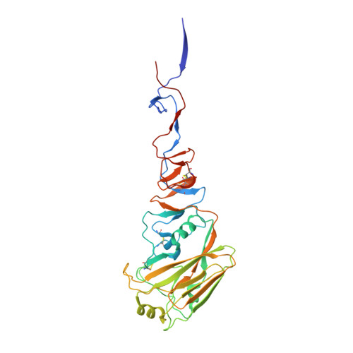
    Antigenic variation among circulating H5N1 highly pathogenic avian influenza A viruses mandates the continuous production of strain-specific pre-pandemic vaccine candidates and represents a significant challenge for pandemic preparedness. Here we assessed the structural, antigenic and receptor-binding properties of three H5N1 HPAI virus hemagglutinins, which were recently selected by the WHO as vaccine candidates [A/Egypt/N03072/2010 (Egypt10, clade 2.2.1), A/Hubei/1/2010 (Hubei10, clade 2.3.2.1) and A/Anhui/1/2005 (Anhui05, clade 2.3.4)]. These analyses revealed that antigenic diversity among these three isolates was restricted to changes in the size and charge of amino acid side chains at a handful of positions, spatially equivalent to the antigenic sites identified in H1 subtype viruses circulating among humans. All three of the H5N1 viruses analyzed in this study were responsible for fatal human infections, with the most recently-isolated strains, Hubei10 and Egypt10, containing multiple residues in the receptor-binding site of the HA, which were suspected to enhance mammalian transmission. However, glycan-binding analyses demonstrated a lack of binding to human α2-6-linked sialic acid receptor analogs for all three HAs, reinforcing the notion that receptor-binding specificity contributes only partially to transmissibility and pathogenesis of HPAI viruses and suggesting that changes in host specificity must be interpreted in the context of the host and environmental factors, as well as the virus as a whole. Together, our data reveal structural linkages with phylogenetic and antigenic analyses of recently emerged H5N1 virus clades and should assist in interpreting the significance of future changes in antigenic and receptor-binding properties.


  • Organizational Affiliation

    Influenza Division, Centers for Disease Control and Prevention, Atlanta, Georgia, United States of America.


Macromolecules
Find similar proteins by:  (by identity cutoff)  |  3D Structure
Entity ID: 1
MoleculeChains Sequence LengthOrganismDetailsImage
Hemagglutinin
A, C, E, G
329Influenza A virus (A/reassortant/IDCDC_RG29(Egypt/N03072/2010 x Puerto Rico/8/1934)(H5N1))Mutation(s): 0 
Gene Names: HA
UniProt
Find proteins for G1JUF7 (Influenza A virus)
Explore G1JUF7 
Go to UniProtKB:  G1JUF7
Entity Groups  
Sequence Clusters30% Identity50% Identity70% Identity90% Identity95% Identity100% Identity
UniProt GroupG1JUF7
Sequence Annotations
Expand
  • Reference Sequence
Find similar proteins by:  (by identity cutoff)  |  3D Structure
Entity ID: 2
MoleculeChains Sequence LengthOrganismDetailsImage
Hemagglutinin
B, D, F, H
181Influenza A virus (A/reassortant/IDCDC_RG29(Egypt/N03072/2010 x Puerto Rico/8/1934)(H5N1))Mutation(s): 0 
Gene Names: HA
UniProt
Find proteins for G1JUF7 (Influenza A virus)
Explore G1JUF7 
Go to UniProtKB:  G1JUF7
Entity Groups  
Sequence Clusters30% Identity50% Identity70% Identity90% Identity95% Identity100% Identity
UniProt GroupG1JUF7
Sequence Annotations
Expand
  • Reference Sequence
Oligosaccharides

Help

Entity ID: 3
MoleculeChains Length2D Diagram Glycosylation3D Interactions
alpha-D-mannopyranose-(1-3)-[alpha-D-mannopyranose-(1-6)]alpha-D-mannopyranose-(1-4)-2-acetamido-2-deoxy-beta-D-glucopyranose-(1-4)-2-acetamido-2-deoxy-beta-D-glucopyranose
I, K
5N-Glycosylation
Glycosylation Resources
GlyTouCan:  G21381MC
GlyCosmos:  G21381MC
GlyGen:  G21381MC
Entity ID: 4
MoleculeChains Length2D Diagram Glycosylation3D Interactions
2-acetamido-2-deoxy-beta-D-glucopyranose-(1-4)-2-acetamido-2-deoxy-beta-D-glucopyranose
J, L, M, N, O
J, L, M, N, O, P
2N-Glycosylation
Glycosylation Resources
GlyTouCan:  G42666HT
GlyCosmos:  G42666HT
GlyGen:  G42666HT
Small Molecules
Ligands 2 Unique
IDChains Name / Formula / InChI Key2D Diagram3D Interactions
NAG
Query on NAG

Download Ideal Coordinates CCD File 
Q [auth A],
T [auth C],
W [auth E],
Z [auth G]
2-acetamido-2-deoxy-beta-D-glucopyranose
C8 H15 N O6
OVRNDRQMDRJTHS-FMDGEEDCSA-N
PO4
Query on PO4

Download Ideal Coordinates CCD File 
AA [auth G]
BA [auth H]
R [auth A]
S [auth B]
U [auth C]
AA [auth G],
BA [auth H],
R [auth A],
S [auth B],
U [auth C],
V [auth D],
X [auth E],
Y [auth F]
PHOSPHATE ION
O4 P
NBIIXXVUZAFLBC-UHFFFAOYSA-K
Experimental Data & Validation

Experimental Data

  • Method: X-RAY DIFFRACTION
  • Resolution: 2.50 Å
  • R-Value Free: 0.227 
  • R-Value Work: 0.193 
  • R-Value Observed: 0.194 
  • Space Group: H 3
Unit Cell:
Length ( Å )Angle ( ˚ )
a = 98.066α = 90
b = 98.066β = 90
c = 655.022γ = 120
Software Package:
Software NamePurpose
HKL-2000data collection
PHASERphasing
REFMACrefinement
HKL-2000data reduction
HKL-2000data scaling

Structure Validation

View Full Validation Report


Ligand Structure Quality Assessment 


Entry History 

Deposition Data

Revision History  (Full details and data files)

  • Version 1.0: 2014-06-25
    Type: Initial release
  • Version 2.0: 2020-07-29
    Type: Remediation
    Reason: Carbohydrate remediation
    Changes: Advisory, Atomic model, Data collection, Database references, Derived calculations, Structure summary