4KVJ

Crystal structure of Oryza sativa fatty acid alpha-dioxygenase with hydrogen peroxide


Experimental Data Snapshot

  • Method: X-RAY DIFFRACTION
  • Resolution: 2.12 Å
  • R-Value Free: 0.208 
  • R-Value Work: 0.170 
  • R-Value Observed: 0.172 

wwPDB Validation   3D Report Full Report


Ligand Structure Quality Assessment 


This is version 1.4 of the entry. See complete history


Literature

Crystal structures of alpha-dioxygenase from Oryza sativa: Insights into substrate binding and activation by hydrogen peroxide.

Zhu, G.Koszelak-Rosenblum, M.Malkowski, M.G.

(2013) Protein Sci 22: 1432-1438

  • DOI: https://doi.org/10.1002/pro.2327
  • Primary Citation of Related Structures:  
    4KVJ, 4KVK, 4KVL

  • PubMed Abstract: 

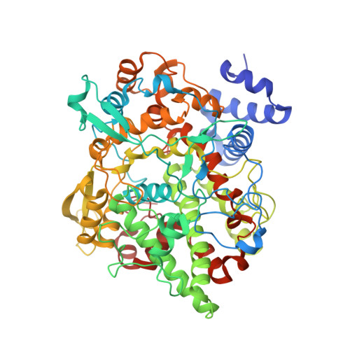
    α-Dioxygenases (α-DOX) are heme-containing enzymes found predominantly in plants and fungi, where they generate oxylipins in response to pathogen attack. α-DOX oxygenate a variety of 14-20 carbon fatty acids containing up to three unsaturated bonds through stereoselective removal of the pro-R hydrogen from the α-carbon by a tyrosyl radical generated via the oxidation of the heme moiety by hydrogen peroxide (H2 O2 ). We determined the X-ray crystal structures of wild type α-DOX from Oryza sativa, the wild type enzyme in complex with H2 O2 , and the catalytically inactive Y379F mutant in complex with the fatty acid palmitic acid (PA). PA binds within the active site cleft of α-DOX such that the carboxylate forms ionic interactions with His-311 and Arg-559. Thr-316 aids in the positioning of carbon-2 for hydrogen abstraction. Twenty-five of the twenty eight contacts made between PA and residues lining the active site occur within the carboxylate and first eight carbons, indicating that interactions within this region of the substrate are responsible for governing selectivity. Comparison of the wild type and H2 O2 structures provides insight into enzyme activation. The binding of H2 O2 at the distal face of the heme displaces residues His-157, Asp-158, and Trp-159 ≈ 2.5 Å from their positions in the wild type structure. As a result, the Oδ2 atom of Asp-158 interacts with the Ca atom in the calcium binding loop, the side chains of Trp-159 and Trp-213 reorient, and the guanidinium group of Arg-559 is repositioned near Tyr-379, poised to interact with the carboxylate group of the substrate.


  • Organizational Affiliation

    Hauptman-Woodward Medical Research Institute, Buffalo, New York.


Macromolecules
Find similar proteins by:  (by identity cutoff)  |  3D Structure
Entity ID: 1
MoleculeChains Sequence LengthOrganismDetailsImage
Fatty acid alpha-oxidase621Oryza sativaMutation(s): 0 
UniProt
Find proteins for Q2QRV3 (Oryza sativa subsp. japonica)
Explore Q2QRV3 
Go to UniProtKB:  Q2QRV3
Entity Groups  
Sequence Clusters30% Identity50% Identity70% Identity90% Identity95% Identity100% Identity
UniProt GroupQ2QRV3
Sequence Annotations
Expand
  • Reference Sequence
Small Molecules
Ligands 10 Unique
IDChains Name / Formula / InChI Key2D Diagram3D Interactions
HEM
Query on HEM

Download Ideal Coordinates CCD File 
B [auth A]PROTOPORPHYRIN IX CONTAINING FE
C34 H32 Fe N4 O4
KABFMIBPWCXCRK-RGGAHWMASA-L
DMU
Query on DMU

Download Ideal Coordinates CCD File 
C [auth A]DECYL-BETA-D-MALTOPYRANOSIDE
C22 H42 O11
WOQQAWHSKSSAGF-WXFJLFHKSA-N
P33
Query on P33

Download Ideal Coordinates CCD File 
F [auth A]3,6,9,12,15,18-HEXAOXAICOSANE-1,20-DIOL
C14 H30 O8
XPJRQAIZZQMSCM-UHFFFAOYSA-N
PG4
Query on PG4

Download Ideal Coordinates CCD File 
D [auth A],
H [auth A]
TETRAETHYLENE GLYCOL
C8 H18 O5
UWHCKJMYHZGTIT-UHFFFAOYSA-N
PGE
Query on PGE

Download Ideal Coordinates CCD File 
G [auth A]TRIETHYLENE GLYCOL
C6 H14 O4
ZIBGPFATKBEMQZ-UHFFFAOYSA-N
PEG
Query on PEG

Download Ideal Coordinates CCD File 
E [auth A]
I [auth A]
J [auth A]
K [auth A]
L [auth A]
E [auth A],
I [auth A],
J [auth A],
K [auth A],
L [auth A],
M [auth A],
N [auth A]
DI(HYDROXYETHYL)ETHER
C4 H10 O3
MTHSVFCYNBDYFN-UHFFFAOYSA-N
CA
Query on CA

Download Ideal Coordinates CCD File 
O [auth A]CALCIUM ION
Ca
BHPQYMZQTOCNFJ-UHFFFAOYSA-N
CL
Query on CL

Download Ideal Coordinates CCD File 
P [auth A],
Q [auth A],
R [auth A],
S [auth A]
CHLORIDE ION
Cl
VEXZGXHMUGYJMC-UHFFFAOYSA-M
PEO
Query on PEO

Download Ideal Coordinates CCD File 
T [auth A]HYDROGEN PEROXIDE
H2 O2
MHAJPDPJQMAIIY-UHFFFAOYSA-N
NA
Query on NA

Download Ideal Coordinates CCD File 
U [auth A],
V [auth A]
SODIUM ION
Na
FKNQFGJONOIPTF-UHFFFAOYSA-N
Experimental Data & Validation

Experimental Data

  • Method: X-RAY DIFFRACTION
  • Resolution: 2.12 Å
  • R-Value Free: 0.208 
  • R-Value Work: 0.170 
  • R-Value Observed: 0.172 
  • Space Group: I 2 2 2
Unit Cell:
Length ( Å )Angle ( ˚ )
a = 72.279α = 90
b = 129.65β = 90
c = 187.711γ = 90
Software Package:
Software NamePurpose
JDirectordata collection
PHASERphasing
REFMACrefinement
autoPROCdata scaling
XDSdata reduction
SCALAdata scaling

Structure Validation

View Full Validation Report


Ligand Structure Quality Assessment 


Entry History 

Deposition Data

Revision History  (Full details and data files)

  • Version 1.0: 2013-08-21
    Type: Initial release
  • Version 1.1: 2013-08-28
    Changes: Database references
  • Version 1.2: 2013-10-16
    Changes: Database references
  • Version 1.3: 2017-11-15
    Changes: Refinement description
  • Version 1.4: 2024-02-28
    Changes: Data collection, Database references, Derived calculations, Refinement description, Structure summary