4KQ6

Product complex of lumazine synthase from candida glabrata


Experimental Data Snapshot

  • Method: X-RAY DIFFRACTION
  • Resolution: 2.24 Å
  • R-Value Free: 0.252 
  • R-Value Work: 0.200 
  • R-Value Observed: 0.201 

wwPDB Validation   3D Report Full Report


Ligand Structure Quality Assessment 


This is version 1.2 of the entry. See complete history


Literature

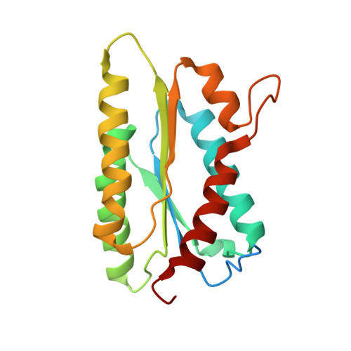
Catalysis product captured in lumazine synthase from the fungal pathogen Candida glabrata.

Shankar, M.Wilbanks, S.M.Nakatani, Y.Monk, B.C.Tyndall, J.D.

(2013) Acta Crystallogr D Biol Crystallogr 69: 1580-1586

  • DOI: https://doi.org/10.1107/S0907444913010949
  • Primary Citation of Related Structures:  
    4KQ6

  • PubMed Abstract: 

    Candida glabrata has emerged as an important fungal pathogen with intrinsic resistance to azole drugs. The limited efficacy of and resistance to existing antifungals is driving the need to identify new drug targets. The enzyme 6,7-dimethyl-8-(D-ribityl)lumazine synthase is part of the riboflavin-biosynthesis pathway essential to fungi and bacteria and is a potential drug target for the development of broad-spectrum antifungal drugs. The X-ray crystal structure of recombinant lumazine synthase from C. glabrata was obtained at 2.24 Å resolution and revealed a dimer of homopentamers, with one in five subunits containing a product molecule from the catalytic reaction.


  • Organizational Affiliation

    School of Pharmacy, University of Otago, PO Box 56, Dunedin 9054, New Zealand.


Macromolecules
Find similar proteins by:  (by identity cutoff)  |  3D Structure
Entity ID: 1
MoleculeChains Sequence LengthOrganismDetailsImage
6,7-dimethyl-8-ribityllumazine synthase
A, B, C, D, E
A, B, C, D, E, F, G, H, I, J
179Nakaseomyces glabratus CBS 138Mutation(s): 0 
Gene Names: CAGL0B01419gRIB4
EC: 2.5.1.78
UniProt
Find proteins for Q6FXA8 (Candida glabrata (strain ATCC 2001 / BCRC 20586 / JCM 3761 / NBRC 0622 / NRRL Y-65 / CBS 138))
Explore Q6FXA8 
Go to UniProtKB:  Q6FXA8
Entity Groups  
Sequence Clusters30% Identity50% Identity70% Identity90% Identity95% Identity100% Identity
UniProt GroupQ6FXA8
Sequence Annotations
Expand
  • Reference Sequence
Small Molecules
Ligands 3 Unique
IDChains Name / Formula / InChI Key2D Diagram3D Interactions
DLZ
Query on DLZ

Download Ideal Coordinates CCD File 
KA [auth I],
R [auth C]
1-deoxy-1-(6,7-dimethyl-2,4-dioxo-3,4-dihydropteridin-8(2H)-yl)-D-ribitol
C13 H18 N4 O6
SXDXRJZUAJBNFL-XKSSXDPKSA-N
SO4
Query on SO4

Download Ideal Coordinates CCD File 
AA [auth F]
BA [auth G]
CA [auth G]
DA [auth G]
FA [auth H]
AA [auth F],
BA [auth G],
CA [auth G],
DA [auth G],
FA [auth H],
GA [auth H],
HA [auth I],
IA [auth I],
K [auth A],
L [auth A],
LA [auth J],
M [auth B],
MA [auth J],
N [auth B],
NA [auth J],
O [auth C],
P [auth C],
Q [auth C],
S [auth D],
T [auth D],
V [auth E],
W [auth E],
Y [auth F],
Z [auth F]
SULFATE ION
O4 S
QAOWNCQODCNURD-UHFFFAOYSA-L
GOL
Query on GOL

Download Ideal Coordinates CCD File 
EA [auth G],
JA [auth I],
OA [auth J],
U [auth D],
X [auth E]
GLYCEROL
C3 H8 O3
PEDCQBHIVMGVHV-UHFFFAOYSA-N
Experimental Data & Validation

Experimental Data

  • Method: X-RAY DIFFRACTION
  • Resolution: 2.24 Å
  • R-Value Free: 0.252 
  • R-Value Work: 0.200 
  • R-Value Observed: 0.201 
  • Space Group: P 21 21 21
Unit Cell:
Length ( Å )Angle ( ˚ )
a = 84.75α = 90
b = 84.84β = 90
c = 310.2γ = 90
Software Package:
Software NamePurpose
PHASERphasing
REFMACrefinement
MOSFLMdata reduction
SCALAdata scaling

Structure Validation

View Full Validation Report


Ligand Structure Quality Assessment 


Entry History 

Deposition Data

Revision History  (Full details and data files)

  • Version 1.0: 2013-05-29
    Type: Initial release
  • Version 1.1: 2013-08-28
    Changes: Database references
  • Version 1.2: 2023-09-20
    Changes: Data collection, Database references, Derived calculations, Refinement description