4KI1

Primitive triclinic crystal form of the human IgE-Fc(epsilon)3-4 bound to its B cell receptor derCD23


Experimental Data Snapshot

  • Method: X-RAY DIFFRACTION
  • Resolution: 3.20 Å
  • R-Value Free: 0.266 
  • R-Value Work: 0.233 
  • R-Value Observed: 0.234 

wwPDB Validation   3D Report Full Report


This is version 2.1 of the entry. See complete history


Literature

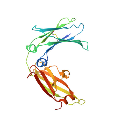
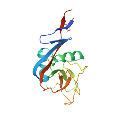
A range of C3-C4 interdomain angles in IgE Fc accommodate binding to its receptor CD23.

Dhaliwal, B.Pang, M.O.Yuan, D.Beavil, A.J.Sutton, B.J.

(2014) Acta Crystallogr Sect F Struct Biol Cryst Commun 70: 305-309

  • DOI: https://doi.org/10.1107/S2053230X14003355
  • Primary Citation of Related Structures:  
    4KI1

  • PubMed Abstract: 

    The antibody IgE plays a central role in allergic disease, functioning principally through two cell-surface receptors: FcℇRI and CD23. FcℇRI on mast cells and basophils mediates the immediate hypersensitivity response, whilst the interaction of IgE with CD23 on B cells regulates IgE production. Crystal structures of the lectin-like `head' domain of CD23 alone and bound to a subfragment of IgE consisting of the dimer of Cℇ3 and Cℇ4 domains (Fcℇ3-4) have recently been determined, revealing flexibility in the IgE-binding site of CD23. Here, a new crystal form of the CD23-Fcℇ3-4 complex with different molecular-packing constraints is reported, which together with the earlier results demonstrates that conformational variability at the interface extends additionally to the IgE Fc and the quaternary structure of its domains.


  • Organizational Affiliation

    Randall Division of Cell and Molecular Biophysics, King's College London, New Hunt's House, Guy's Campus, London SE1 1UL, England.


Macromolecules
Find similar proteins by:  (by identity cutoff)  |  3D Structure
Entity ID: 1
MoleculeChains Sequence LengthOrganismDetailsImage
IG EPSILON CHAIN C REGION
A, B, C, D
223Homo sapiensMutation(s): 2 
Gene Names: IGHE
UniProt & NIH Common Fund Data Resources
Find proteins for P01854 (Homo sapiens)
Explore P01854 
Go to UniProtKB:  P01854
PHAROS:  P01854
GTEx:  ENSG00000211891 
Entity Groups  
Sequence Clusters30% Identity50% Identity70% Identity90% Identity95% Identity100% Identity
UniProt GroupP01854
Sequence Annotations
Expand
  • Reference Sequence
Find similar proteins by:  (by identity cutoff)  |  3D Structure
Entity ID: 2
MoleculeChains Sequence LengthOrganismDetailsImage
LOW AFFINITY IMMUNOGLOBULIN EPSILON FC RECEPTOR
E, F, G, H
143Homo sapiensMutation(s): 0 
Gene Names: FCER2CD23ACLEC4JFCE2IGEBF
UniProt & NIH Common Fund Data Resources
Find proteins for P06734 (Homo sapiens)
Explore P06734 
Go to UniProtKB:  P06734
PHAROS:  P06734
GTEx:  ENSG00000104921 
Entity Groups  
Sequence Clusters30% Identity50% Identity70% Identity90% Identity95% Identity100% Identity
UniProt GroupP06734
Sequence Annotations
Expand
  • Reference Sequence
Oligosaccharides

Help

Entity ID: 3
MoleculeChains Length2D Diagram Glycosylation3D Interactions
alpha-D-mannopyranose-(1-3)-[alpha-D-mannopyranose-(1-6)]beta-D-mannopyranose-(1-4)-2-acetamido-2-deoxy-beta-D-glucopyranose-(1-4)-2-acetamido-2-deoxy-beta-D-glucopyranose
I, J, K, L
5N-Glycosylation
Glycosylation Resources
GlyTouCan:  G22768VO
GlyCosmos:  G22768VO
GlyGen:  G22768VO
Experimental Data & Validation

Experimental Data

  • Method: X-RAY DIFFRACTION
  • Resolution: 3.20 Å
  • R-Value Free: 0.266 
  • R-Value Work: 0.233 
  • R-Value Observed: 0.234 
  • Space Group: P 1
Unit Cell:
Length ( Å )Angle ( ˚ )
a = 48.79α = 100.67
b = 63.84β = 90.13
c = 163.89γ = 103.49
Software Package:
Software NamePurpose
MOSFLMdata reduction
PHASESphasing
BUSTERrefinement
SCALAdata scaling

Structure Validation

View Full Validation Report


Entry History 

Deposition Data

Revision History  (Full details and data files)

  • Version 1.0: 2014-03-05
    Type: Initial release
  • Version 1.1: 2014-03-12
    Changes: Database references
  • Version 1.2: 2014-09-24
    Changes: Database references
  • Version 2.0: 2020-07-29
    Type: Remediation
    Reason: Carbohydrate remediation
    Changes: Atomic model, Data collection, Database references, Derived calculations, Structure summary
  • Version 2.1: 2023-09-20
    Changes: Data collection, Database references, Refinement description, Structure summary