4KGE

Crystal structure of near-infrared fluorescent protein with an extended stokes shift, pH 4.5


Experimental Data Snapshot

  • Method: X-RAY DIFFRACTION
  • Resolution: 2.30 Å
  • R-Value Free: 0.232 
  • R-Value Work: 0.188 
  • R-Value Observed: 0.190 

wwPDB Validation   3D Report Full Report


This is version 1.4 of the entry. See complete history


Literature

Extended Stokes shift in fluorescent proteins: chromophore-protein interactions in a near-infrared TagRFP675 variant.

Piatkevich, K.D.Malashkevich, V.N.Morozova, K.S.Nemkovich, N.A.Almo, S.C.Verkhusha, V.V.

(2013) Sci Rep 3: 1847-1847

  • DOI: https://doi.org/10.1038/srep01847
  • Primary Citation of Related Structures:  
    4KGE, 4KGF

  • PubMed Abstract: 

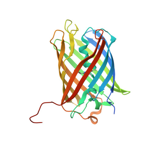
    Most GFP-like fluorescent proteins exhibit small Stokes shifts (10-45 nm) due to rigidity of the chromophore environment that excludes non-fluorescent relaxation to a ground state. An unusual near-infrared derivative of the red fluorescent protein mKate, named TagRFP675, exhibits the Stokes shift, which is 30 nm extended comparing to that of the parental protein. In physiological conditions, TagRFP675 absorbs at 598 nm and emits at 675 nm that makes it the most red-shifted protein of the GFP-like protein family. In addition, its emission maximum strongly depends on the excitation wavelength. Structures of TagRFP675 revealed the common DsRed-like chromophore, which, however, interacts with the protein matrix via an extensive network of hydrogen bonds capable of large flexibility. Based on the spectroscopic, biochemical, and structural analysis we suggest that the rearrangement of the hydrogen bond interactions between the chromophore and the protein matrix is responsible for the TagRFP675 spectral properties.


  • Organizational Affiliation

    Department of Anatomy and Structural Biology and Gruss-Lipper Biophotonics Center, Bronx, New York 10461, United States.


Macromolecules
Find similar proteins by:  (by identity cutoff)  |  3D Structure
Entity ID: 1
MoleculeChains Sequence LengthOrganismDetailsImage
TagRFP675, red fluorescent protein
A, B
243synthetic constructMutation(s): 0 
Entity Groups  
Sequence Clusters30% Identity50% Identity70% Identity90% Identity95% Identity100% Identity
Sequence Annotations
Expand
  • Reference Sequence
Small Molecules
Modified Residues  1 Unique
IDChains TypeFormula2D DiagramParent
NRQ
Query on NRQ
A, B
L-PEPTIDE LINKINGC16 H17 N3 O4 SMET, TYR, GLY
Experimental Data & Validation

Experimental Data

  • Method: X-RAY DIFFRACTION
  • Resolution: 2.30 Å
  • R-Value Free: 0.232 
  • R-Value Work: 0.188 
  • R-Value Observed: 0.190 
  • Space Group: P 61 2 2
Unit Cell:
Length ( Å )Angle ( ˚ )
a = 106.05α = 90
b = 106.05β = 90
c = 219.526γ = 120
Software Package:
Software NamePurpose
SCALEPACKdata scaling
PHASERphasing
REFMACrefinement
PDB_EXTRACTdata extraction
CBASSdata collection
HKL-2000data reduction

Structure Validation

View Full Validation Report


Entry History 

Revision History  (Full details and data files)

  • Version 1.0: 2013-05-08
    Type: Initial release
  • Version 1.1: 2014-01-08
    Changes: Database references
  • Version 1.2: 2018-01-24
    Changes: Structure summary
  • Version 1.3: 2023-09-20
    Changes: Data collection, Database references, Derived calculations, Refinement description
  • Version 1.4: 2023-12-06
    Changes: Data collection