


Crystal structure of the mitochondrial peroxiredoxin from Leishmania braziliensis in the dimeric form
Souza, T.A.C.B., Morais, M.A.B., Giuseppe, P.O., Murakami, M.T.To be published.
Experimental Data Snapshot
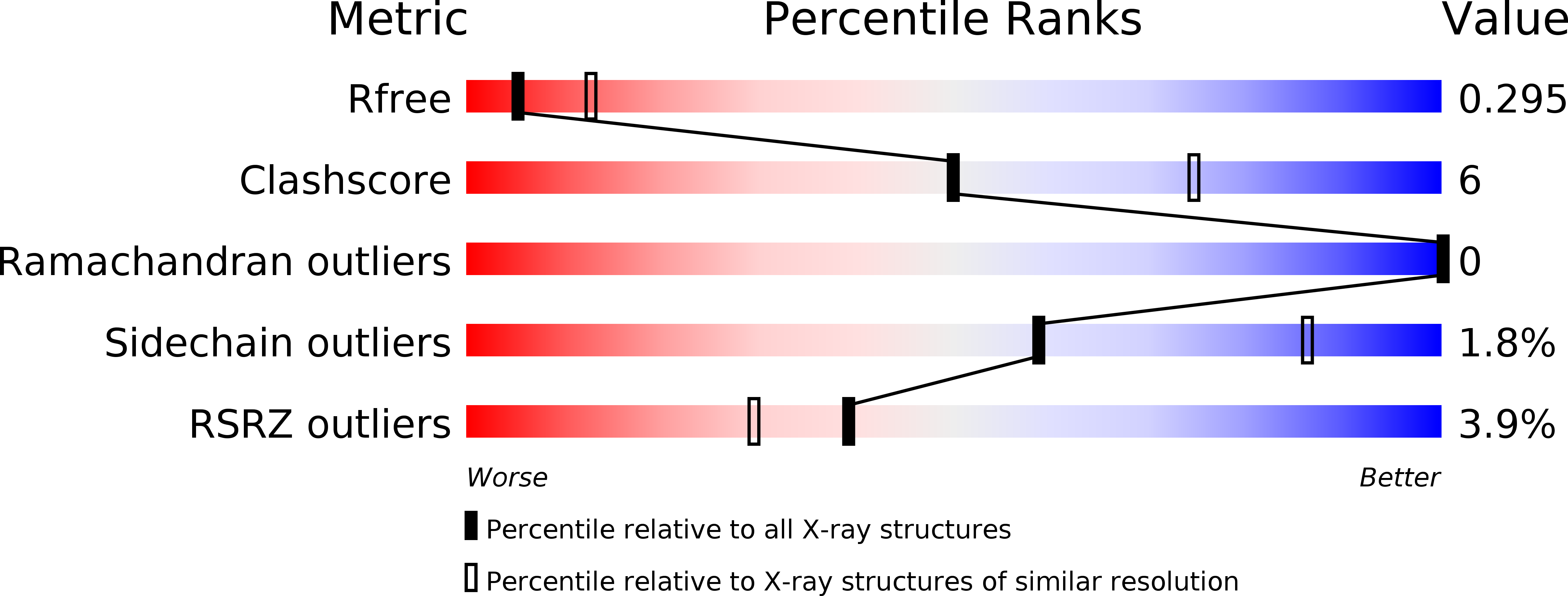
wwPDB Validation 3D Report Full Report
Entity ID: 1 | |||||
|---|---|---|---|---|---|
| Molecule | Chains | Sequence Length | Organism | Details | Image |
| Peroxidoxin | 213 | Leishmania braziliensis | Mutation(s): 0 Gene Names: LBRM_23_0050 EC: 1.11.1 | 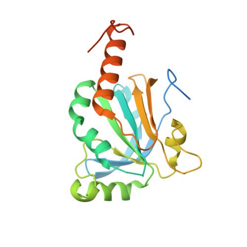 | |
UniProt | |||||
Find proteins for A4HCL7 (Leishmania braziliensis) Explore A4HCL7 Go to UniProtKB: A4HCL7 | |||||
Entity Groups | |||||
| Sequence Clusters | 30% Identity50% Identity70% Identity90% Identity95% Identity100% Identity | ||||
| UniProt Group | A4HCL7 | ||||
Sequence AnnotationsExpand | |||||
| |||||
| Length ( Å ) | Angle ( ˚ ) |
|---|---|
| a = 132.485 | α = 90 |
| b = 132.485 | β = 90 |
| c = 44.627 | γ = 90 |
| Software Name | Purpose |
|---|---|
| XSCALE | data scaling |
| MOLREP | phasing |
| PHENIX | refinement |
| PDB_EXTRACT | data extraction |
RCSB PDB (citation) is hosted by
RCSB PDB is a member of the

