4KC3

Cytokine/receptor binary complex


Experimental Data Snapshot

  • Method: X-RAY DIFFRACTION
  • Resolution: 3.27 Å
  • R-Value Free: 0.283 
  • R-Value Work: 0.238 
  • R-Value Observed: 0.241 

wwPDB Validation   3D Report Full Report


Ligand Structure Quality Assessment 


This is version 1.5 of the entry. See complete history


Literature

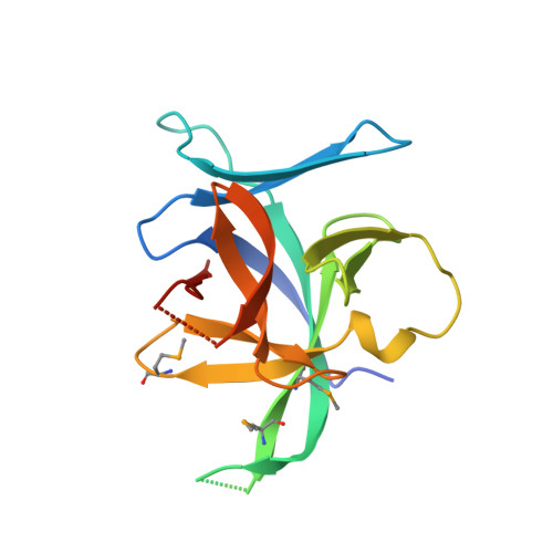
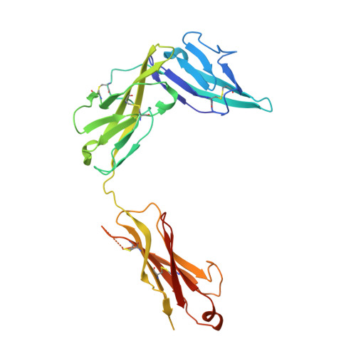
Structural insights into the interaction of IL-33 with its receptors.

Liu, X.Hammel, M.He, Y.Tainer, J.A.Jeng, U.S.Zhang, L.Wang, S.Wang, X.

(2013) Proc Natl Acad Sci U S A 110: 14918-14923

  • DOI: https://doi.org/10.1073/pnas.1308651110
  • Primary Citation of Related Structures:  
    4KC3

  • PubMed Abstract: 

    Interleukin (IL)-33 is an important member of the IL-1 family that has pleiotropic activities in innate and adaptive immune responses in host defense and disease. It signals through its ligand-binding primary receptor ST2 and IL-1 receptor accessory protein (IL-1RAcP), both of which are members of the IL-1 receptor family. To clarify the interaction of IL-33 with its receptors, we determined the crystal structure of IL-33 in complex with the ectodomain of ST2 at a resolution of 3.27 Å. Coupled with structure-based mutagenesis and binding assay, the structural results define the molecular mechanism by which ST2 specifically recognizes IL-33. Structural comparison with other ligand-receptor complexes in the IL-1 family indicates that surface-charge complementarity is critical in determining ligand-binding specificity of IL-1 primary receptors. Combined crystallography and small-angle X-ray-scattering studies reveal that ST2 possesses hinge flexibility between the D3 domain and D1D2 module, whereas IL-1RAcP exhibits a rigid conformation in the unbound state in solution. The molecular flexibility of ST2 provides structural insights into domain-level conformational change of IL-1 primary receptors upon ligand binding, and the rigidity of IL-1RAcP explains its inability to bind ligands directly. The solution architecture of IL-33-ST2-IL-1RAcP complex from small-angle X-ray-scattering analysis resembles IL-1β-IL-1RII-IL-1RAcP and IL-1β-IL-1RI-IL-1RAcP crystal structures. The collective results confer IL-33 structure-function relationships, supporting and extending a general model for ligand-receptor assembly and activation in the IL-1 family.


  • Organizational Affiliation

    Ministry of Education Key Laboratory of Protein Science, Center for Structural Biology, School of Life Sciences, Tsinghua University, Beijing 100084, China.


Macromolecules
Find similar proteins by:  (by identity cutoff)  |  3D Structure
Entity ID: 1
MoleculeChains Sequence LengthOrganismDetailsImage
Interleukin-33159Homo sapiensMutation(s): 0 
Gene Names: IL33C9orf26IL1F11NFHEV
UniProt & NIH Common Fund Data Resources
Find proteins for O95760 (Homo sapiens)
Explore O95760 
Go to UniProtKB:  O95760
PHAROS:  O95760
GTEx:  ENSG00000137033 
Entity Groups  
Sequence Clusters30% Identity50% Identity70% Identity90% Identity95% Identity100% Identity
UniProt GroupO95760
Sequence Annotations
Expand
  • Reference Sequence
Find similar proteins by:  (by identity cutoff)  |  3D Structure
Entity ID: 2
MoleculeChains Sequence LengthOrganismDetailsImage
Interleukin-1 receptor-like 1309Homo sapiensMutation(s): 0 
Gene Names: IL1RL1DER4ST2T1
UniProt & NIH Common Fund Data Resources
Find proteins for Q01638 (Homo sapiens)
Explore Q01638 
Go to UniProtKB:  Q01638
PHAROS:  Q01638
GTEx:  ENSG00000115602 
Entity Groups  
Sequence Clusters30% Identity50% Identity70% Identity90% Identity95% Identity100% Identity
UniProt GroupQ01638
Sequence Annotations
Expand
  • Reference Sequence
Small Molecules
Modified Residues  1 Unique
IDChains TypeFormula2D DiagramParent
MSE
Query on MSE
A
L-PEPTIDE LINKINGC5 H11 N O2 SeMET
Experimental Data & Validation

Experimental Data

  • Method: X-RAY DIFFRACTION
  • Resolution: 3.27 Å
  • R-Value Free: 0.283 
  • R-Value Work: 0.238 
  • R-Value Observed: 0.241 
  • Space Group: H 3 2
Unit Cell:
Length ( Å )Angle ( ˚ )
a = 329.924α = 90
b = 329.924β = 90
c = 43.731γ = 120
Software Package:
Software NamePurpose
HKL-2000data collection
PHASERphasing
PHENIXrefinement
HKL-2000data reduction
HKL-2000data scaling

Structure Validation

View Full Validation Report


Ligand Structure Quality Assessment 


Entry History 

Deposition Data

Revision History  (Full details and data files)

  • Version 1.0: 2013-08-28
    Type: Initial release
  • Version 1.1: 2013-09-11
    Changes: Database references
  • Version 1.2: 2013-09-25
    Changes: Database references
  • Version 1.3: 2020-07-29
    Type: Remediation
    Reason: Carbohydrate remediation
    Changes: Data collection, Database references, Derived calculations, Structure summary
  • Version 1.4: 2023-09-20
    Changes: Data collection, Database references, Refinement description, Structure summary
  • Version 1.5: 2023-12-06
    Changes: Data collection