4K2J

Decameric ring structure of KSHV (HHV-8) latency-associated nuclear antigen (LANA) DNA binding domain


Experimental Data Snapshot

  • Method: X-RAY DIFFRACTION
  • Resolution: 2.05 Å
  • R-Value Free: 0.226 
  • R-Value Work: 0.182 
  • R-Value Observed: 0.184 

wwPDB Validation   3D Report Full Report


This is version 1.1 of the entry. See complete history


Literature

Molecular Basis for Oligomeric-DNA Binding and Episome Maintenance by KSHV LANA.

Domsic, J.F.Chen, H.S.Lu, F.Marmorstein, R.Lieberman, P.M.

(2013) PLoS Pathog 9: e1003672-e1003672

  • DOI: https://doi.org/10.1371/journal.ppat.1003672
  • Primary Citation of Related Structures:  
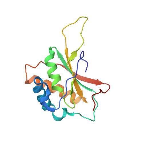
    4K2J

  • PubMed Abstract: 

    LANA is the KSHV-encoded terminal repeat binding protein essential for viral replication and episome maintenance during latency. We have determined the X-ray crystal structure of LANA C-terminal DNA binding domain (LANADBD) to reveal its capacity to form a decameric ring with an exterior DNA binding surface. The dimeric core is structurally similar to EBV EBNA1 with an N-terminal arm that regulates DNA binding and is required for replication function. The oligomeric interface between LANA dimers is dispensable for single site DNA binding, but is required for cooperative DNA binding, replication function, and episome maintenance. We also identify a basic patch opposite of the DNA binding surface that is responsible for the interaction with BRD proteins and contributes to episome maintenance function. The structural features of LANADBD suggest a novel mechanism of episome maintenance through DNA-binding induced oligomeric assembly.


  • Organizational Affiliation

    Gene Expression and Regulation Program, The Wistar Institute, Philadelphia, Pennsylvania, United States of America.


Macromolecules
Find similar proteins by:  (by identity cutoff)  |  3D Structure
Entity ID: 1
MoleculeChains Sequence LengthOrganismDetailsImage
KSHV (HHV-8) latency-associated nuclear antigen (LANA)
A, B, C, D, E
A, B, C, D, E, F, G, H, I, J
140Human gammaherpesvirus 8Mutation(s): 0 
Gene Names: ORF73
UniProt
Find proteins for Q9DUN0 (Human herpesvirus 8)
Explore Q9DUN0 
Go to UniProtKB:  Q9DUN0
Entity Groups  
Sequence Clusters30% Identity50% Identity70% Identity90% Identity95% Identity100% Identity
UniProt GroupQ9DUN0
Sequence Annotations
Expand
  • Reference Sequence
Small Molecules
Ligands 2 Unique
IDChains Name / Formula / InChI Key2D Diagram3D Interactions
FMT
Query on FMT

Download Ideal Coordinates CCD File 
DA [auth G]
IA [auth I]
K [auth A]
NA [auth J]
OA [auth J]
DA [auth G],
IA [auth I],
K [auth A],
NA [auth J],
OA [auth J],
P [auth C],
Q [auth C],
U [auth E],
V [auth E]
FORMIC ACID
C H2 O2
BDAGIHXWWSANSR-UHFFFAOYSA-N
CL
Query on CL

Download Ideal Coordinates CCD File 
AA [auth F]
BA [auth F]
CA [auth F]
EA [auth G]
FA [auth H]
AA [auth F],
BA [auth F],
CA [auth F],
EA [auth G],
FA [auth H],
GA [auth H],
HA [auth H],
JA [auth I],
KA [auth I],
L [auth A],
LA [auth I],
M [auth A],
MA [auth I],
N [auth B],
O [auth B],
PA [auth J],
R [auth C],
S [auth C],
T [auth D],
W [auth E],
X [auth E],
Y [auth E],
Z [auth E]
CHLORIDE ION
Cl
VEXZGXHMUGYJMC-UHFFFAOYSA-M
Experimental Data & Validation

Experimental Data

  • Method: X-RAY DIFFRACTION
  • Resolution: 2.05 Å
  • R-Value Free: 0.226 
  • R-Value Work: 0.182 
  • R-Value Observed: 0.184 
  • Space Group: P 1 21 1
Unit Cell:
Length ( Å )Angle ( ˚ )
a = 51.439α = 90
b = 175.176β = 95.3
c = 97.065γ = 90
Software Package:
Software NamePurpose
CBASSdata collection
PHASERphasing
PHENIXrefinement
HKL-3000data reduction
HKL-3000data scaling

Structure Validation

View Full Validation Report


Entry History 

Deposition Data

Revision History  (Full details and data files)

  • Version 1.0: 2013-11-06
    Type: Initial release
  • Version 1.1: 2024-02-28
    Changes: Data collection, Database references, Derived calculations