4JOE

CFTR Associated Ligand (CAL) PDZ domain bound to peptide A-iCAL36 (ANSRAPTSII)


Experimental Data Snapshot

  • Method: X-RAY DIFFRACTION
  • Resolution: 1.14 Å
  • R-Value Free: 0.195 
  • R-Value Work: 0.182 
  • R-Value Observed: 0.185 

wwPDB Validation   3D Report Full Report


This is version 1.1 of the entry. See complete history


Literature

Stereochemical Preferences Modulate Affinity and Selectivity among Five PDZ Domains that Bind CFTR: Comparative Structural and Sequence Analyses.

Amacher, J.F.Cushing, P.R.Brooks, L.Boisguerin, P.Madden, D.R.

(2014) Structure 22: 82-93

  • DOI: https://doi.org/10.1016/j.str.2013.09.019
  • Primary Citation of Related Structures:  
    4JOE, 4JOF, 4JOG, 4JOH, 4JOJ, 4JOK, 4JOP, 4JOR, 4K6Y, 4K72, 4K75, 4K76, 4K78

  • PubMed Abstract: 

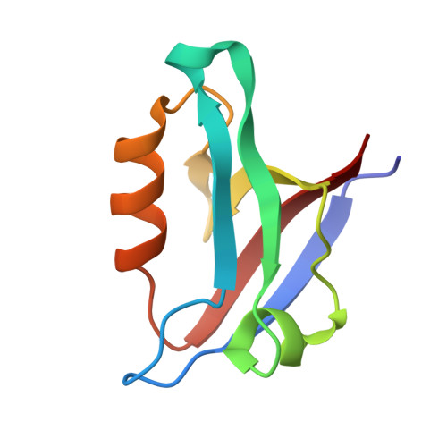
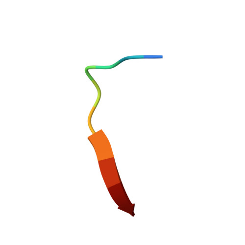
    PDZ domain interactions are involved in signaling and trafficking pathways that coordinate crucial cellular processes. Alignment-based PDZ binding motifs identify the few most favorable residues at certain positions along the peptide backbone. However, sequences that bind the CAL (CFTR-associated ligand) PDZ domain reveal only a degenerate motif that overpredicts the true number of high-affinity interactors. Here, we combine extended peptide-array motif analysis with biochemical techniques to show that non-motif "modulator" residues influence CAL binding. The crystallographic structures of 13 CAL:peptide complexes reveal defined, but accommodating stereochemical environments at non-motif positions, which are reflected in modulator preferences uncovered by multisequence substitutional arrays. These preferences facilitate the identification of high-affinity CAL binding sequences and differentially affect CAL and NHERF PDZ binding. As a result, they also help determine the specificity of a PDZ domain network that regulates the trafficking of CFTR at the apical membrane.


  • Organizational Affiliation

    Department of Biochemistry, Geisel School of Medicine at Dartmouth, Hanover, NH 03755, USA.


Macromolecules
Find similar proteins by:  (by identity cutoff)  |  3D Structure
Entity ID: 1
MoleculeChains Sequence LengthOrganismDetailsImage
Golgi-associated PDZ and coiled-coil motif-containing protein
A, B
87Homo sapiensMutation(s): 0 
Gene Names: GOPCCALFIG
UniProt & NIH Common Fund Data Resources
Find proteins for Q9HD26 (Homo sapiens)
Explore Q9HD26 
Go to UniProtKB:  Q9HD26
PHAROS:  Q9HD26
GTEx:  ENSG00000047932 
Entity Groups  
Sequence Clusters30% Identity50% Identity70% Identity90% Identity95% Identity100% Identity
UniProt GroupQ9HD26
Sequence Annotations
Expand
  • Reference Sequence

Find similar proteins by:  Sequence   |   3D Structure  

Entity ID: 2
MoleculeChains Sequence LengthOrganismDetailsImage
A-iCAL36 peptide
C, D
10N/AMutation(s): 0 
Entity Groups  
Sequence Clusters30% Identity50% Identity70% Identity90% Identity95% Identity100% Identity
Sequence Annotations
Expand
  • Reference Sequence
Experimental Data & Validation

Experimental Data

  • Method: X-RAY DIFFRACTION
  • Resolution: 1.14 Å
  • R-Value Free: 0.195 
  • R-Value Work: 0.182 
  • R-Value Observed: 0.185 
  • Space Group: P 21 21 21
Unit Cell:
Length ( Å )Angle ( ˚ )
a = 36.679α = 90
b = 47.816β = 90
c = 97.665γ = 90
Software Package:
Software NamePurpose
HKL-2000data collection
PHENIXmodel building
PHENIXrefinement
XDSdata reduction
XSCALEdata scaling
PHENIXphasing

Structure Validation

View Full Validation Report


Entry History 

Deposition Data

Revision History  (Full details and data files)

  • Version 1.0: 2014-01-22
    Type: Initial release
  • Version 1.1: 2023-09-20
    Changes: Data collection, Database references, Derived calculations, Refinement description