4JJ2

High resolution structure of a C-terminal fragment of the T4 phage gp5 beta-helix


Experimental Data Snapshot

  • Method: X-RAY DIFFRACTION
  • Resolution: 1.28 Å
  • R-Value Free: 0.178 
  • R-Value Work: 0.153 
  • R-Value Observed: 0.154 

wwPDB Validation   3D Report Full Report


Ligand Structure Quality Assessment 


This is version 1.2 of the entry. See complete history


Literature

Structure and Biophysical Properties of a Triple-Stranded Beta-Helix Comprising the Central Spike of Bacteriophage T4.

Buth, S.A.Menin, L.Shneider, M.M.Engel, J.Boudko, S.P.Leiman, P.G.

(2015) Viruses 7: 4676-4706

  • DOI: https://doi.org/10.3390/v7082839
  • Primary Citation of Related Structures:  
    4JJ2, 4OSD

  • PubMed Abstract: 

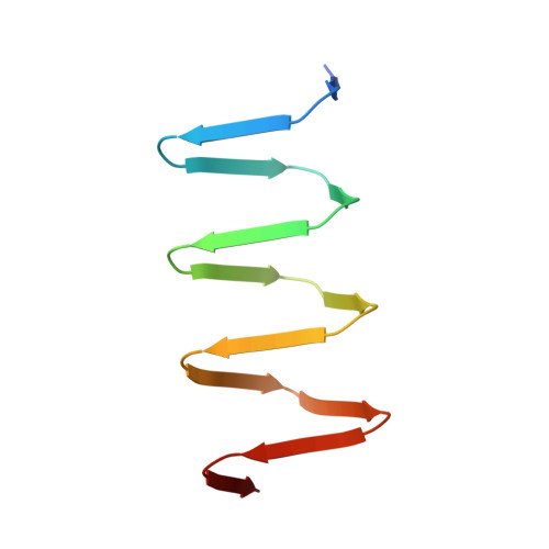
    Gene product 5 (gp5) of bacteriophage T4 is a spike-shaped protein that functions to disrupt the membrane of the target cell during phage infection. Its C-terminal domain is a long and slender β-helix that is formed by three polypeptide chains wrapped around a common symmetry axis akin to three interdigitated corkscrews. The folding and biophysical properties of such triple-stranded β-helices, which are topologically related to amyloid fibers, represent an unsolved biophysical problem. Here, we report structural and biophysical characterization of T4 gp5 β-helix and its truncated mutants of different lengths. A soluble fragment that forms a dimer of trimers and that could comprise a minimal self-folding unit has been identified. Surprisingly, the hydrophobic core of the β-helix is small. It is located near the C-terminal end of the β-helix and contains a centrally positioned and hydrated magnesium ion. A large part of the β-helix interior comprises a large elongated cavity that binds palmitic, stearic, and oleic acids in an extended conformation suggesting that these molecules might participate in the folding of the complete β-helix.


  • Organizational Affiliation

    Institute of Physics of Biological Systems, École Polytechnique Fédérale de Lausanne (EPFL), BSP 415, 1015 Lausanne, Switzerland. sergii.buth@epfl.ch.


Macromolecules
Find similar proteins by:  (by identity cutoff)  |  3D Structure
Entity ID: 1
MoleculeChains Sequence LengthOrganismDetailsImage
Tail-associated lysozyme
A, B, C
95Tequatrovirus T4Mutation(s): 0 
Gene Names: 5
EC: 3.2.1.17
UniProt
Find proteins for P16009 (Enterobacteria phage T4)
Explore P16009 
Go to UniProtKB:  P16009
Entity Groups  
Sequence Clusters30% Identity50% Identity70% Identity90% Identity95% Identity100% Identity
UniProt GroupP16009
Sequence Annotations
Expand
  • Reference Sequence
Experimental Data & Validation

Experimental Data

  • Method: X-RAY DIFFRACTION
  • Resolution: 1.28 Å
  • R-Value Free: 0.178 
  • R-Value Work: 0.153 
  • R-Value Observed: 0.154 
  • Space Group: C 2 2 21
Unit Cell:
Length ( Å )Angle ( ˚ )
a = 57.609α = 90
b = 72.764β = 90
c = 130.49γ = 90
Software Package:
Software NamePurpose
SHELXmodel building
SHELXrefinement
DENZOdata reduction
SCALEPACKdata scaling
SHELXphasing

Structure Validation

View Full Validation Report


Ligand Structure Quality Assessment 


Entry History 

Deposition Data

Revision History  (Full details and data files)

  • Version 1.0: 2014-03-12
    Type: Initial release
  • Version 1.1: 2017-11-15
    Changes: Refinement description
  • Version 1.2: 2023-02-08
    Changes: Database references, Derived calculations