4J88

Dark-state structure of sfGFP containing the unnatural amino acid p-azido-phenylalanine at residue 66


Experimental Data Snapshot

  • Method: X-RAY DIFFRACTION
  • Resolution: 2.08 Å
  • R-Value Free: 0.228 
  • R-Value Work: 0.165 
  • R-Value Observed: 0.168 

wwPDB Validation   3D Report Full Report


This is version 2.0 of the entry. See complete history


Literature

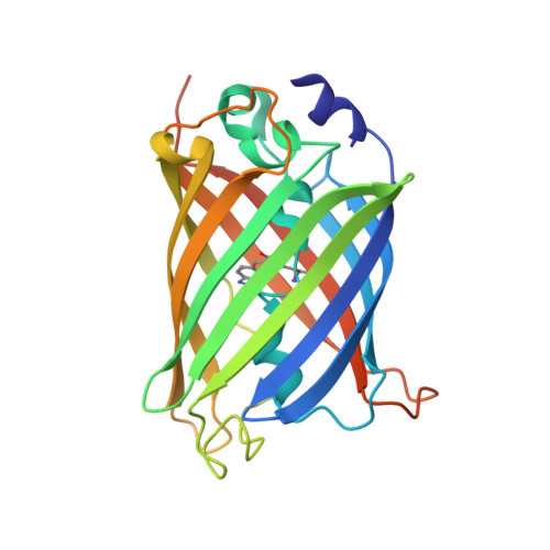
Different Photochemical Events of a Genetically Encoded Phenyl Azide Define and Modulate GFP Fluorescence.

Reddington, S.C.Rizkallah, P.J.Watson, P.D.Pearson, R.Tippmann, E.M.Jones, D.D.

(2013) Angew Chem Int Ed Engl 52: 5974-5977

  • DOI: https://doi.org/10.1002/anie.201301490
  • Primary Citation of Related Structures:  
    4J88, 4J89, 4J8A

  • PubMed Abstract: 

    Expanding the genetic code opens new avenues to modulate protein function in real time. By genetically incorporating photoreactive phenyl azide, the fluorescent properties of green fluorescent protein (GFP) can be modulated by light. Depending on the residue in GFP programmed to incorporate the phenyl azide, different effects on function and photochemical pathways are observed.


  • Organizational Affiliation

    School of Biosciences, Cardiff University, Cardiff, UK.


Macromolecules
Find similar proteins by:  (by identity cutoff)  |  3D Structure
Entity ID: 1
MoleculeChains Sequence LengthOrganismDetailsImage
Green fluorescent protein
A, B
245Aequorea victoriaMutation(s): 10 
Gene Names: GFP
UniProt
Find proteins for P42212 (Aequorea victoria)
Explore P42212 
Go to UniProtKB:  P42212
Entity Groups  
Sequence Clusters30% Identity50% Identity70% Identity90% Identity95% Identity100% Identity
UniProt GroupP42212
Sequence Annotations
Expand
  • Reference Sequence
Small Molecules
Ligands 3 Unique
IDChains Name / Formula / InChI Key2D Diagram3D Interactions
TRS
Query on TRS

Download Ideal Coordinates CCD File 
AA [auth A],
BA [auth A],
Z [auth A]
2-AMINO-2-HYDROXYMETHYL-PROPANE-1,3-DIOL
C4 H12 N O3
LENZDBCJOHFCAS-UHFFFAOYSA-O
SO4
Query on SO4

Download Ideal Coordinates CCD File 
C [auth A]
CA [auth B]
D [auth A]
DA [auth B]
E [auth A]
C [auth A],
CA [auth B],
D [auth A],
DA [auth B],
E [auth A],
EA [auth B],
F [auth A],
FA [auth B],
GA [auth B]
SULFATE ION
O4 S
QAOWNCQODCNURD-UHFFFAOYSA-L
EDO
Query on EDO

Download Ideal Coordinates CCD File 
G [auth A]
H [auth A]
HA [auth B]
I [auth A]
IA [auth B]
G [auth A],
H [auth A],
HA [auth B],
I [auth A],
IA [auth B],
J [auth A],
JA [auth B],
K [auth A],
KA [auth B],
L [auth A],
LA [auth B],
M [auth A],
N [auth A],
O [auth A],
P [auth A],
Q [auth A],
R [auth A],
S [auth A],
T [auth A],
U [auth A],
V [auth A],
W [auth A],
X [auth A],
Y [auth A]
1,2-ETHANEDIOL
C2 H6 O2
LYCAIKOWRPUZTN-UHFFFAOYSA-N
Modified Residues  1 Unique
IDChains TypeFormula2D DiagramParent
CQ1
Query on CQ1
A, B
PEPTIDE LINKINGC15 H16 N6 O4THR, TYR, GLY
Experimental Data & Validation

Experimental Data

  • Method: X-RAY DIFFRACTION
  • Resolution: 2.08 Å
  • R-Value Free: 0.228 
  • R-Value Work: 0.165 
  • R-Value Observed: 0.168 
  • Space Group: P 21 21 21
Unit Cell:
Length ( Å )Angle ( ˚ )
a = 52.43α = 90
b = 97.56β = 90
c = 102.9γ = 90
Software Package:
Software NamePurpose
SCALAdata scaling
PHASERphasing
REFMACrefinement
PDB_EXTRACTdata extraction
GDAdata collection
xia2data reduction

Structure Validation

View Full Validation Report


Entry History 

Deposition Data

Revision History  (Full details and data files)

  • Version 1.0: 2013-06-26
    Type: Initial release
  • Version 1.1: 2023-09-20
    Changes: Data collection, Database references, Derived calculations, Refinement description
  • Version 2.0: 2023-11-15
    Changes: Atomic model, Data collection, Derived calculations