4IB3

Structure of cAMP dependent protein kinase A in complex with ADP, phosphorylated peptide pSP20, and no metal


Experimental Data Snapshot

  • Method: X-RAY DIFFRACTION
  • Resolution: 2.20 Å
  • R-Value Free: 0.228 
  • R-Value Work: 0.182 

wwPDB Validation   3D Report Full Report


Ligand Structure Quality Assessment 


This is version 1.2 of the entry. See complete history


Literature

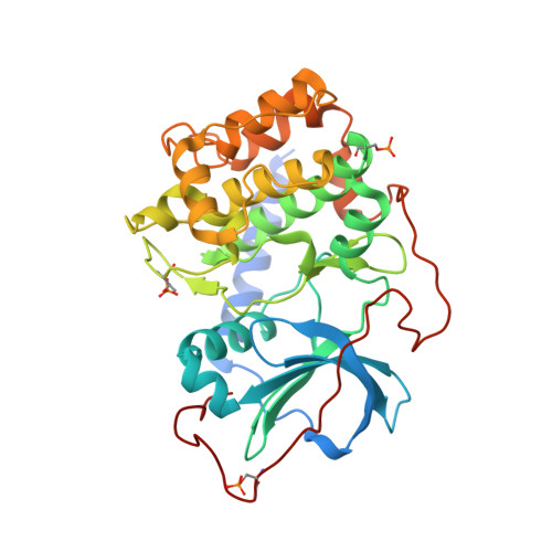
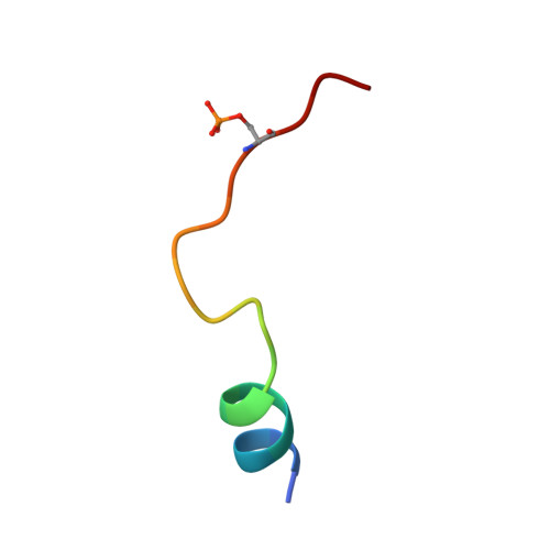
Metal-Free cAMP-Dependent Protein Kinase Can Catalyze Phosphoryl Transfer.

Gerlits, O.Das, A.Keshwani, M.M.Taylor, S.Waltman, M.J.Langan, P.Heller, W.T.Kovalevsky, A.

(2014) Biochemistry 53: 3179-3186

  • DOI: https://doi.org/10.1021/bi5000965
  • Primary Citation of Related Structures:  
    4IB0, 4IB1, 4IB3, 4O21, 4O22

  • PubMed Abstract: 

    X-ray structures of several ternary product complexes of the catalytic subunit of cAMP-dependent protein kinase (PKAc) have been determined with no bound metal ions and with Na(+) or K(+) coordinated at two metal-binding sites. The metal-free PKAc and the enzyme with alkali metals were able to facilitate the phosphoryl transfer reaction. In all studied complexes, the ATP and the substrate peptide (SP20) were modified into the products ADP and the phosphorylated peptide. The products of the phosphotransfer reaction were also found when ATP-γS, a nonhydrolyzable ATP analogue, reacted with SP20 in the PKAc active site containing no metals. Single turnover enzyme kinetics measurements utilizing (32)P-labeled ATP confirmed the phosphotransferase activity of the enzyme in the absence of metal ions and in the presence of alkali metals. In addition, the structure of the apo-PKAc binary complex with SP20 suggests that the sequence of binding events may become ordered in a metal-free environment, with SP20 binding first to prime the enzyme for subsequent ATP binding. Comparison of these structures reveals conformational and hydrogen bonding changes that might be important for the mechanism of catalysis.


  • Organizational Affiliation

    Biology and Soft Matter Division, Oak Ridge National Laboratory , Oak Ridge, Tennessee 37831, United States.


Macromolecules
Find similar proteins by:  (by identity cutoff)  |  3D Structure
Entity ID: 1
MoleculeChains Sequence LengthOrganismDetailsImage
cAMP-dependent protein kinase catalytic subunit alpha356Mus musculusMutation(s): 0 
Gene Names: PrkacaPkaca
EC: 2.7.11.11
UniProt
Find proteins for P05132 (Mus musculus)
Explore P05132 
Go to UniProtKB:  P05132
Entity Groups  
Sequence Clusters30% Identity50% Identity70% Identity90% Identity95% Identity100% Identity
UniProt GroupP05132
Sequence Annotations
Expand
  • Reference Sequence

Find similar proteins by:  Sequence   |   3D Structure  

Entity ID: 2
MoleculeChains Sequence LengthOrganismDetailsImage
phosphorylated pseudo-substrate peptide pSP20B [auth S]20Homo sapiensMutation(s): 0 
UniProt & NIH Common Fund Data Resources
Find proteins for P61925 (Homo sapiens)
Explore P61925 
Go to UniProtKB:  P61925
PHAROS:  P61925
GTEx:  ENSG00000171033 
Entity Groups  
Sequence Clusters30% Identity50% Identity70% Identity90% Identity95% Identity100% Identity
UniProt GroupP61925
Sequence Annotations
Expand
  • Reference Sequence
Small Molecules
Ligands 1 Unique
IDChains Name / Formula / InChI Key2D Diagram3D Interactions
ADP
Query on ADP

Download Ideal Coordinates CCD File 
C [auth A]ADENOSINE-5'-DIPHOSPHATE
C10 H15 N5 O10 P2
XTWYTFMLZFPYCI-KQYNXXCUSA-N
Modified Residues  2 Unique
IDChains TypeFormula2D DiagramParent
SEP
Query on SEP
A
L-PEPTIDE LINKINGC3 H8 N O6 PSER
TPO
Query on TPO
A
L-PEPTIDE LINKINGC4 H10 N O6 PTHR
Experimental Data & Validation

Experimental Data

  • Method: X-RAY DIFFRACTION
  • Resolution: 2.20 Å
  • R-Value Free: 0.228 
  • R-Value Work: 0.182 
  • Space Group: P 21 21 21
Unit Cell:
Length ( Å )Angle ( ˚ )
a = 57.541α = 90
b = 79.309β = 90
c = 98.418γ = 90
Software Package:
Software NamePurpose
HKL-3000data collection
CNSrefinement
HKL-3000data reduction
HKL-3000data scaling
CNSphasing

Structure Validation

View Full Validation Report


Ligand Structure Quality Assessment 


Entry History 

Deposition Data

Revision History  (Full details and data files)

  • Version 1.0: 2013-12-11
    Type: Initial release
  • Version 1.1: 2014-05-21
    Changes: Database references
  • Version 1.2: 2014-05-28
    Changes: Database references