4I3G

Crystal Structure of DesR, a beta-glucosidase from Streptomyces venezuelae in complex with D-glucose.


Experimental Data Snapshot

  • Method: X-RAY DIFFRACTION
  • Resolution: 1.40 Å
  • R-Value Free: 0.199 
  • R-Value Work: 0.172 
  • R-Value Observed: 0.173 

wwPDB Validation   3D Report Full Report


Ligand Structure Quality Assessment 


This is version 1.3 of the entry. See complete history


Literature

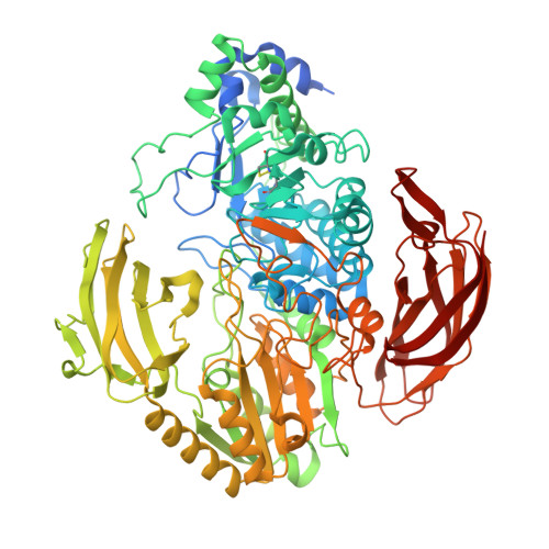
The structure of DesR from Streptomyces venezuelae, a beta-glucosidase involved in macrolide activation.

Zmudka, M.W.Thoden, J.B.Holden, H.M.

(2013) Protein Sci 22: 883-892

  • DOI: https://doi.org/10.1002/pro.2204
  • Primary Citation of Related Structures:  
    4I3G

  • PubMed Abstract: 

    Antibiotics have, indeed, altered the course of human history as is evidenced by the increase in human life expectancy since the 1940s. Many of these natural compounds are produced by bacteria that, by necessity, must have efficient self-resistance mechanisms. The methymycin/pikromycin producing species Streptomyces venezuelae, for example, utilizes β-glucosylation of its macrolide products to neutralize their effects within the confines of the cell. Once released into the environment, these compounds are activated by the removal of the glucose moiety. In S. venezuelae, the enzyme responsible for removal of the sugar from the parent compound is encoded by the desR gene and referred to as DesR. It is a secreted enzyme containing 828 amino acid residues, and it is known to be a retaining glycosidase. Here, we describe the structure of the DesR/D-glucose complex determined to 1.4-Å resolution. The overall architecture of the enzyme can be envisioned in terms of three regions: a catalytic core and two auxiliary domains. The catalytic core harbors the binding platform for the glucose ligand. The first auxiliary domain adopts a "PA14 fold," whereas the second auxiliary domain contains an immunoglobulin-like fold. Asp 273 and Glu 578 are in the proper orientation to function as the catalytic base and proton donor, respectively, required for catalysis. The overall fold of the core region places DesR into the GH3 glycoside hydrolase family of enzymes. Comparison of the DesR structure with the β-glucosidase from Kluyveromyces marxianus shows that their PA14 domains assume remarkably different orientations.


  • Organizational Affiliation

    Department of Biochemistry, University of Wisconsin, Madison, Wisconsin 53706, USA.


Macromolecules
Find similar proteins by:  (by identity cutoff)  |  3D Structure
Entity ID: 1
MoleculeChains Sequence LengthOrganismDetailsImage
Beta-glucosidase
A, B
829Streptomyces venezuelaeMutation(s): 0 
Gene Names: desR
UniProt
Find proteins for C5IXN7 (Streptomyces venezuelae)
Explore C5IXN7 
Go to UniProtKB:  C5IXN7
Entity Groups  
Sequence Clusters30% Identity50% Identity70% Identity90% Identity95% Identity100% Identity
UniProt GroupC5IXN7
Sequence Annotations
Expand
  • Reference Sequence
Small Molecules
Ligands 4 Unique
IDChains Name / Formula / InChI Key2D Diagram3D Interactions
MPO
Query on MPO

Download Ideal Coordinates CCD File 
L [auth A],
T [auth B]
3[N-MORPHOLINO]PROPANE SULFONIC ACID
C7 H15 N O4 S
DVLFYONBTKHTER-UHFFFAOYSA-N
BGC
Query on BGC

Download Ideal Coordinates CCD File 
C [auth A],
O [auth B]
beta-D-glucopyranose
C6 H12 O6
WQZGKKKJIJFFOK-VFUOTHLCSA-N
SO4
Query on SO4

Download Ideal Coordinates CCD File 
D [auth A]
E [auth A]
F [auth A]
G [auth A]
H [auth A]
D [auth A],
E [auth A],
F [auth A],
G [auth A],
H [auth A],
I [auth A],
J [auth A],
K [auth A],
P [auth B],
Q [auth B],
R [auth B],
S [auth B]
SULFATE ION
O4 S
QAOWNCQODCNURD-UHFFFAOYSA-L
EDO
Query on EDO

Download Ideal Coordinates CCD File 
M [auth A],
N [auth A],
U [auth B],
V [auth B]
1,2-ETHANEDIOL
C2 H6 O2
LYCAIKOWRPUZTN-UHFFFAOYSA-N
Experimental Data & Validation

Experimental Data

  • Method: X-RAY DIFFRACTION
  • Resolution: 1.40 Å
  • R-Value Free: 0.199 
  • R-Value Work: 0.172 
  • R-Value Observed: 0.173 
  • Space Group: P 21 21 2
Unit Cell:
Length ( Å )Angle ( ˚ )
a = 131.269α = 90
b = 198.314β = 90
c = 67.023γ = 90
Software Package:
Software NamePurpose
HKL-3000data collection
PHASERphasing
REFMACrefinement
HKL-3000data reduction
HKL-3000data scaling

Structure Validation

View Full Validation Report


Ligand Structure Quality Assessment 


Entry History 

Deposition Data

Revision History  (Full details and data files)

  • Version 1.0: 2013-02-20
    Type: Initial release
  • Version 1.1: 2013-08-28
    Changes: Database references
  • Version 1.2: 2020-07-29
    Type: Remediation
    Reason: Carbohydrate remediation
    Changes: Data collection, Database references, Derived calculations, Structure summary
  • Version 1.3: 2023-09-20
    Changes: Data collection, Database references, Refinement description, Structure summary