


Crystal structure and substrate-binding mode of cellulase Cel5A from a metagenome library
Zhuang, N., Lee, K.H.To be published.
Experimental Data Snapshot
wwPDB Validation 3D Report Full Report

Entity ID: 1 | |||||
|---|---|---|---|---|---|
| Molecule | Chains | Sequence Length | Organism | Details | Image |
| Cellulase | 359 | uncultured bacterium | Mutation(s): 0 | 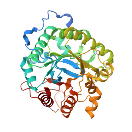 | |
UniProt | |||||
Find proteins for I6PLH5 (uncultured bacterium) Explore I6PLH5 Go to UniProtKB: I6PLH5 | |||||
Entity Groups | |||||
| Sequence Clusters | 30% Identity50% Identity70% Identity90% Identity95% Identity100% Identity | ||||
| UniProt Group | I6PLH5 | ||||
Sequence AnnotationsExpand | |||||
| |||||
| Ligands 1 Unique | |||||
|---|---|---|---|---|---|
| ID | Chains | Name / Formula / InChI Key | 2D Diagram | 3D Interactions | |
| GOL Query on GOL | B [auth A], C [auth A], D [auth A], E [auth A], F [auth A] | GLYCEROL C3 H8 O3 PEDCQBHIVMGVHV-UHFFFAOYSA-N |  | ||
| Modified Residues 1 Unique | |||||
|---|---|---|---|---|---|
| ID | Chains | Type | Formula | 2D Diagram | Parent |
| MSE Query on MSE | A | L-PEPTIDE LINKING | C5 H11 N O2 Se |  | MET |
| Length ( Å ) | Angle ( ˚ ) |
|---|---|
| a = 137.771 | α = 90 |
| b = 137.771 | β = 90 |
| c = 137.771 | γ = 90 |
| Software Name | Purpose |
|---|---|
| HKL-2000 | data collection |
| SOLVE | phasing |
| REFMAC | refinement |
| HKL-2000 | data reduction |
| HKL-2000 | data scaling |
RCSB PDB (citation) is hosted by
RCSB PDB is a member of the

