4HTJ

Crystallographic structure of the membrane-proximal ectodomain of the human receptor-type protein-tyrosine phosphatase phogrin at pH 4.6


Experimental Data Snapshot

  • Method: X-RAY DIFFRACTION
  • Resolution: 2.01 Å
  • R-Value Free: 0.238 
  • R-Value Work: 0.203 
  • R-Value Observed: 0.204 

wwPDB Validation   3D Report Full Report


This is version 1.6 of the entry. See complete history


Literature

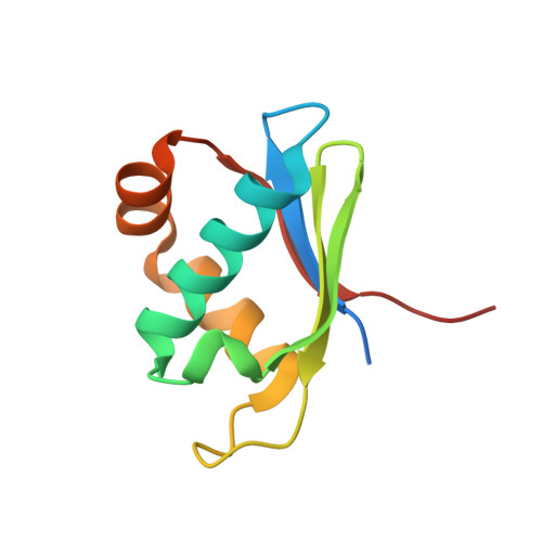
X-ray structure of the mature ectodomain of phogrin.

Noguera, M.E.Primo, M.E.Jakoncic, J.Poskus, E.Solimena, M.Ermacora, M.R.

(2015) J Struct Funct Genomics 16: 1-9

  • DOI: https://doi.org/10.1007/s10969-014-9191-0
  • Primary Citation of Related Structures:  
    4HTI, 4HTJ

  • PubMed Abstract: 

    Phogrin/IA-2β and ICA512/IA-2 are two paralogs receptor-type protein-tyrosine phosphatases (RPTP) that localize in secretory granules of various neuroendocrine cells. In pancreatic islet β-cells, they participate in the regulation of insulin secretion, ensuring proper granulogenesis, and β-cell proliferation. The role of their cytoplasmic tail has been partially unveiled, while that of their luminal region remains unclear. To advance the understanding of its structure-function relationship, the X-ray structure of the mature ectodomain of phogrin (ME phogrin) at pH 7.4 and 4.6 has been solved at 1.95- and 2.01-Å resolution, respectively. Similarly to the ME of ICA512, ME phogrin adopts a ferredoxin-like fold: a sheet of four antiparallel β-strands packed against two α-helices. Sequence conservation among vertebrates, plants and insects suggests that the structural similarity extends to all the receptor family. Crystallized ME phogrin is monomeric, in agreement with solution studies but in striking contrast with the behavior of homodimeric ME ICA512. The structural details that may cause the quaternary structure differences are analyzed. The results provide a basis for building models of the overall orientation and oligomerization state of the receptor in biological membranes.


  • Organizational Affiliation

    Departamento de Ciencia y Tecnología, Universidad Nacional de Quilmes, Sáenz Peña 352, B1876BXD, Bernal, Buenos Aires, Argentina.


Macromolecules
Find similar proteins by:  (by identity cutoff)  |  3D Structure
Entity ID: 1
MoleculeChains Sequence LengthOrganismDetailsImage
Receptor-type tyrosine-protein phosphatase N299Homo sapiensMutation(s): 0 
Gene Names: PTPRN2KIAA0387
EC: 3.1.3.48
UniProt & NIH Common Fund Data Resources
Find proteins for Q92932 (Homo sapiens)
Explore Q92932 
Go to UniProtKB:  Q92932
PHAROS:  Q92932
GTEx:  ENSG00000155093 
Entity Groups  
Sequence Clusters30% Identity50% Identity70% Identity90% Identity95% Identity100% Identity
UniProt GroupQ92932
Sequence Annotations
Expand
  • Reference Sequence
Experimental Data & Validation

Experimental Data

  • Method: X-RAY DIFFRACTION
  • Resolution: 2.01 Å
  • R-Value Free: 0.238 
  • R-Value Work: 0.203 
  • R-Value Observed: 0.204 
  • Space Group: P 61 2 2
Unit Cell:
Length ( Å )Angle ( ˚ )
a = 54.742α = 90
b = 54.742β = 90
c = 149.147γ = 120
Software Package:
Software NamePurpose
DENZOdata reduction
SCALEPACKdata scaling
MOLREPphasing
REFMACrefinement
PDB_EXTRACTdata extraction
HKL-2000data collection
HKL-2000data reduction

Structure Validation

View Full Validation Report


Entry History 

Deposition Data

Revision History  (Full details and data files)

  • Version 1.0: 2012-11-28
    Type: Initial release
  • Version 1.1: 2012-12-19
    Changes: Database references
  • Version 1.2: 2013-03-20
    Changes: Database references
  • Version 1.3: 2013-07-24
    Changes: Database references
  • Version 1.4: 2015-03-18
    Changes: Database references
  • Version 1.5: 2017-11-15
    Changes: Refinement description
  • Version 1.6: 2023-09-20
    Changes: Data collection, Database references, Refinement description