Glycan shifting on hepatitis C virus (HCV) e2 glycoprotein is a mechanism for escape from broadly neutralizing antibodies.
Pantua, H., Diao, J., Ultsch, M., Hazen, M., Mathieu, M., McCutcheon, K., Takeda, K., Date, S., Cheung, T.K., Phung, Q., Hass, P., Arnott, D., Hongo, J.A., Matthews, D.J., Brown, A., Patel, A.H., Kelley, R.F., Eigenbrot, C., Kapadia, S.B.(2013) J Mol Biol 425: 1899-1914
- PubMed: 23458406
- DOI: https://doi.org/10.1016/j.jmb.2013.02.025
- Primary Citation of Related Structures:
4HS6, 4HS8 - PubMed Abstract:
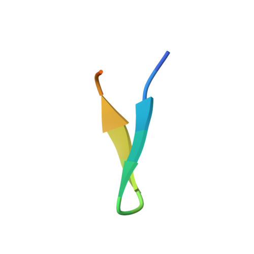
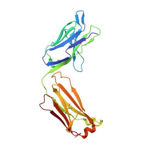
Hepatitis C virus (HCV) infection is a major cause of liver disease and hepatocellular carcinoma. Glycan shielding has been proposed to be a mechanism by which HCV masks broadly neutralizing epitopes on its viral glycoproteins. However, the role of altered glycosylation in HCV resistance to broadly neutralizing antibodies is not fully understood. Here, we have generated potent HCV neutralizing antibodies hu5B3.v3 and MRCT10.v362 that, similar to the previously described AP33 and HCV1, bind to a highly conserved linear epitope on E2. We utilize a combination of in vitro resistance selections using the cell culture infectious HCV and structural analyses to identify mechanisms of HCV resistance to hu5B3.v3 and MRCT10.v362. Ultra deep sequencing from in vitro HCV resistance selection studies identified resistance mutations at asparagine N417 (N417S, N417T and N417G) as early as 5days post treatment. Comparison of the glycosylation status of soluble versions of the E2 glycoprotein containing the respective resistance mutations revealed a glycosylation shift from N417 to N415 in the N417S and N417T E2 proteins. The N417G E2 variant was glycosylated neither at residue 415 nor at residue 417 and remained sensitive to MRCT10.v362. Structural analyses of the E2 epitope bound to hu5B3.v3 Fab and MRCT10.v362 Fab using X-ray crystallography confirmed that residue N415 is buried within the antibody-peptide interface. Thus, in addition to previously described mutations at N415 that abrogate the β-hairpin structure of this E2 linear epitope, we identify a second escape mechanism, termed glycan shifting, that decreases the efficacy of broadly neutralizing HCV antibodies.
Organizational Affiliation:
Department of Infectious Diseases, Genentech, 1 DNA Way, South San Francisco, CA 94080, USA.