4HKL

Crystal Structures of Mutant Endo-beta-1,4-xylanase II Complexed with substrate (1.15 A) and Products (1.6 A)


Experimental Data Snapshot

  • Method: X-RAY DIFFRACTION
  • Resolution: 1.10 Å
  • R-Value Free: 0.129 
  • R-Value Work: 0.119 
  • R-Value Observed: 0.120 

wwPDB Validation   3D Report Full Report


This is version 1.4 of the entry. See complete history


Literature

X-ray crystallographic studies of family 11 xylanase Michaelis and product complexes: implications for the catalytic mechanism.

Wan, Q.Zhang, Q.Hamilton-Brehm, S.Weiss, K.Mustyakimov, M.Coates, L.Langan, P.Graham, D.Kovalevsky, A.

(2014) Acta Crystallogr D Biol Crystallogr 70: 11-23

  • DOI: https://doi.org/10.1107/S1399004713023626
  • Primary Citation of Related Structures:  
    4HK8, 4HK9, 4HKL, 4HKO, 4HKW, 6K9X

  • PubMed Abstract: 

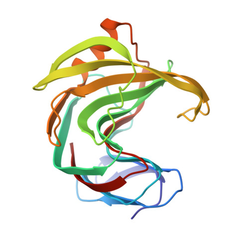
    Xylanases catalyze the hydrolysis of plant hemicellulose xylan into oligosaccharides by cleaving the main-chain glycosidic linkages connecting xylose subunits. To study ligand binding and to understand how the pH constrains the activity of the enzyme, variants of the Trichoderma reesei xylanase were designed to either abolish its activity (E177Q) or to change its pH optimum (N44H). An E177Q-xylohexaose complex structure was obtained at 1.15 Å resolution which represents a pseudo-Michaelis complex and confirmed the conformational movement of the thumb region owing to ligand binding. Co-crystallization of N44H with xylohexaose resulted in a hydrolyzed xylotriose bound in the active site. Co-crystallization of the wild-type enzyme with xylopentaose trapped an aglycone xylotriose and a transglycosylated glycone product. Replacing amino acids near Glu177 decreased the xylanase activity but increased the relative activity at alkaline pH. The substrate distortion in the E177Q-xylohexaose structure expands the possible conformational itinerary of this xylose ring during the enzyme-catalyzed xylan-hydrolysis reaction.


  • Organizational Affiliation

    Biology and Soft Matter Division, Oak Ridge National Laboratory, PO Box 2008, Oak Ridge, TN 37831, USA.


Macromolecules
Find similar proteins by:  (by identity cutoff)  |  3D Structure
Entity ID: 1
MoleculeChains Sequence LengthOrganismDetailsImage
Endo-1,4-beta-xylanase 2189Trichoderma reeseiMutation(s): 1 
Gene Names: xyn2
EC: 3.2.1.8
UniProt
Find proteins for P36217 (Hypocrea jecorina (strain ATCC 56765 / BCRC 32924 / NRRL 11460 / Rut C-30))
Explore P36217 
Go to UniProtKB:  P36217
Entity Groups  
Sequence Clusters30% Identity50% Identity70% Identity90% Identity95% Identity100% Identity
UniProt GroupP36217
Sequence Annotations
Expand
  • Reference Sequence
Experimental Data & Validation

Experimental Data

  • Method: X-RAY DIFFRACTION
  • Resolution: 1.10 Å
  • R-Value Free: 0.129 
  • R-Value Work: 0.119 
  • R-Value Observed: 0.120 
  • Space Group: P 21 21 21
Unit Cell:
Length ( Å )Angle ( ˚ )
a = 48.307α = 90
b = 59.032β = 90
c = 69.717γ = 90
Software Package:
Software NamePurpose
DENZOdata reduction
SCALEPACKdata scaling
PHASERphasing
PHENIXrefinement
PDB_EXTRACTdata extraction
HKL-2000data collection
HKL-2000data reduction

Structure Validation

View Full Validation Report


Entry History 

Deposition Data

Revision History  (Full details and data files)

  • Version 1.0: 2014-01-08
    Type: Initial release
  • Version 1.1: 2014-02-12
    Changes: Database references
  • Version 1.2: 2017-11-15
    Changes: Refinement description
  • Version 1.3: 2018-01-24
    Changes: Structure summary
  • Version 1.4: 2024-02-28
    Changes: Data collection, Database references, Derived calculations