4HAN

Crystal structure of Galectin 8 with NDP52 peptide


Experimental Data Snapshot

  • Method: X-RAY DIFFRACTION
  • Resolution: 2.55 Å
  • R-Value Free: 0.214 
  • R-Value Work: 0.191 
  • R-Value Observed: 0.193 

wwPDB Validation   3D Report Full Report


Ligand Structure Quality Assessment 


This is version 1.3 of the entry. See complete history


Literature

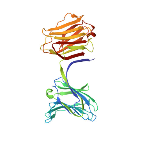
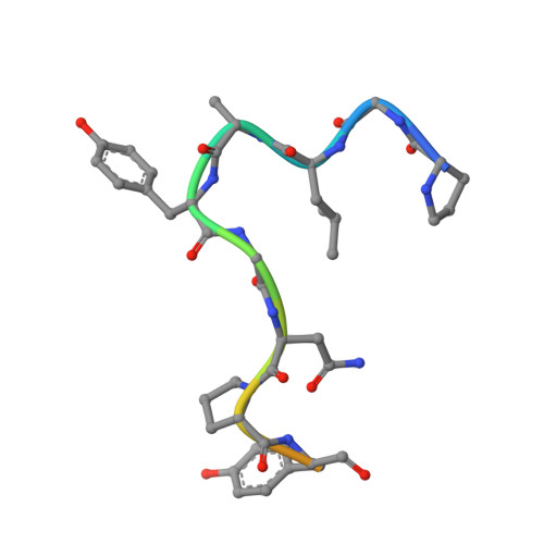
Structural basis for recognition of autophagic receptor NDP52 by the sugar receptor galectin-8.

Kim, B.W.Hong, S.B.Kim, J.H.Kwon, D.H.Song, H.K.

(2013) Nat Commun 4: 1613-1613

  • DOI: https://doi.org/10.1038/ncomms2606
  • Primary Citation of Related Structures:  
    4HAN

  • PubMed Abstract: 

    Infectious bacteria are cleared from mammalian cells by host autophagy in combination with other upstream cellular components, such as the autophagic receptor NDP52 and sugar receptor galectin-8. However, the detailed molecular basis of the interaction between these two receptors remains to be elucidated. Here, we report the biochemical characterization of both NDP52 and galectin-8 as well as the crystal structure of galectin-8 complexed with an NDP52 peptide. The unexpected observation of nicotinamide adenine dinucleotide located at the carbohydrate-binding site expands our knowledge of the sugar-binding specificity of galectin-8. The NDP52-galectin-8 complex structure explains the key determinants for recognition on both receptors and defines a special orientation of N- and C-terminal carbohydrate recognition domains of galectin-8. Dimeric NDP52 forms a ternary complex with two monomeric galectin-8 molecules as well as two LC3C molecules. These results lay the groundwork for understanding how host cells target bacterial pathogens for autophagy.


  • Organizational Affiliation

    Division of Life Sciences, School of Life Sciences and Biotechnology, Korea University, Anam-Dong, Seongbuk-Gu, Seoul, Korea.


Macromolecules
Find similar proteins by:  (by identity cutoff)  |  3D Structure
Entity ID: 1
MoleculeChains Sequence LengthOrganismDetailsImage
Galectin-8
A, B
293Homo sapiensMutation(s): 0 
UniProt & NIH Common Fund Data Resources
Find proteins for O00214 (Homo sapiens)
Explore O00214 
Go to UniProtKB:  O00214
PHAROS:  O00214
GTEx:  ENSG00000116977 
Entity Groups  
Sequence Clusters30% Identity50% Identity70% Identity90% Identity95% Identity100% Identity
UniProt GroupO00214
Sequence Annotations
Expand
  • Reference Sequence

Find similar proteins by:  Sequence   |   3D Structure  

Entity ID: 2
MoleculeChains Sequence LengthOrganismDetailsImage
Calcium-binding and coiled-coil domain-containing protein 2
C, D
14Homo sapiensMutation(s): 0 
Gene Names: CALCOCO2NDP52
UniProt & NIH Common Fund Data Resources
Find proteins for Q13137 (Homo sapiens)
Explore Q13137 
Go to UniProtKB:  Q13137
PHAROS:  Q13137
GTEx:  ENSG00000136436 
Entity Groups  
Sequence Clusters30% Identity50% Identity70% Identity90% Identity95% Identity100% Identity
UniProt GroupQ13137
Sequence Annotations
Expand
  • Reference Sequence
Experimental Data & Validation

Experimental Data

  • Method: X-RAY DIFFRACTION
  • Resolution: 2.55 Å
  • R-Value Free: 0.214 
  • R-Value Work: 0.191 
  • R-Value Observed: 0.193 
  • Space Group: P 31 1 2
Unit Cell:
Length ( Å )Angle ( ˚ )
a = 71.884α = 90
b = 71.884β = 90
c = 314.493γ = 120
Software Package:
Software NamePurpose
HKL-2000data collection
PHENIXrefinement
HKL-2000data reduction
HKL-2000data scaling

Structure Validation

View Full Validation Report


Ligand Structure Quality Assessment 


Entry History 

Deposition Data

Revision History  (Full details and data files)

  • Version 1.0: 2013-03-20
    Type: Initial release
  • Version 1.1: 2013-05-15
    Changes: Database references
  • Version 1.2: 2017-07-26
    Changes: Source and taxonomy
  • Version 1.3: 2024-02-28
    Changes: Data collection, Database references, Derived calculations