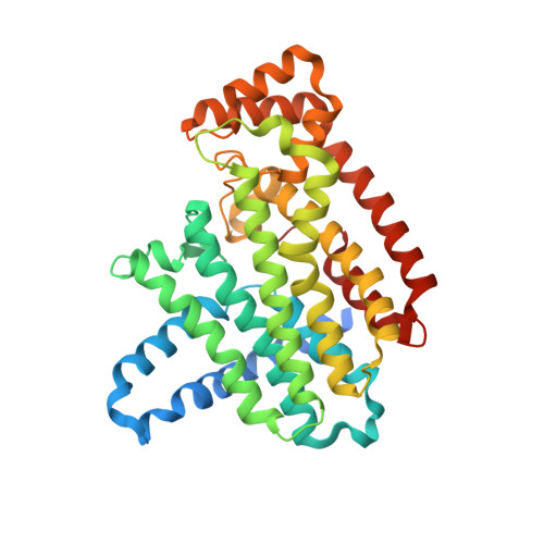
Ternary complex structures of human farnesyl pyrophosphate synthase bound with a novel inhibitor and secondary ligands provide insights into the molecular details of the enzyme's active site closure.
Park, J., Lin, Y.S., De Schutter, J.W., Tsantrizos, Y.S., Berghuis, A.M.(2012) BMC Struct Biol 12: 32-32
- PubMed: 23234314
- DOI: https://doi.org/10.1186/1472-6807-12-32
- Primary Citation of Related Structures:
4H5C, 4H5D, 4H5E - PubMed Abstract:
Human farnesyl pyrophosphate synthase (FPPS) controls intracellular levels of farnesyl pyrophosphate, which is essential for various biological processes. Bisphosphonate inhibitors of human FPPS are valuable therapeutics for the treatment of bone-resorption disorders and have also demonstrated efficacy in multiple tumor types. Inhibition of human FPPS by bisphosphonates in vivo is thought to involve closing of the enzyme's C-terminal tail induced by the binding of the second substrate isopentenyl pyrophosphate (IPP). This conformational change, which occurs through a yet unclear mechanism, seals off the enzyme's active site from the solvent environment and is essential for catalysis. The crystal structure of human FPPS in complex with a novel bisphosphonate YS0470 and in the absence of a second substrate showed partial ordering of the tail in the closed conformation. We have determined crystal structures of human FPPS in ternary complex with YS0470 and the secondary ligands inorganic phosphate (Pi), inorganic pyrophosphate (PPi), and IPP. Binding of PPi or IPP to the enzyme-inhibitor complex, but not that of Pi, resulted in full ordering of the C-terminal tail, which is most notably characterized by the anchoring of the R351 side chain to the main frame of the enzyme. Isothermal titration calorimetry experiments demonstrated that PPi binds more tightly to the enzyme-inhibitor complex than IPP, and differential scanning fluorometry experiments confirmed that Pi binding does not induce the tail ordering. Structure analysis identified a cascade of conformational changes required for the C-terminal tail rigidification involving Y349, F238, and Q242. The residues K57 and N59 upon PPi/IPP binding undergo subtler conformational changes, which may initiate this cascade. In human FPPS, Y349 functions as a safety switch that prevents any futile C-terminal closure and is locked in the "off" position in the absence of bound IPP. Q242 plays the role of a gatekeeper and directly controls the anchoring of R351 side chain. The interactions between the residues K57 and N59 and those upstream and downstream of Y349 are likely responsible for the switch activation. The findings of this study can be exploited for structure-guided optimization of existing inhibitors as well as development of new pharmacophores.
Organizational Affiliation:
Department of Biochemistry, McGill University, Montreal, Canada.