4H2L

Deer mouse hemoglobin in hydrated format


Experimental Data Snapshot

  • Method: X-RAY DIFFRACTION
  • Resolution: 1.78 Å
  • R-Value Free: 0.201 
  • R-Value Work: 0.162 
  • R-Value Observed: 0.165 

wwPDB Validation   3D Report Full Report


Ligand Structure Quality Assessment 


This is version 1.2 of the entry. See complete history


Literature

Deer mouse hemoglobin exhibits a lowered oxygen affinity owing to mobility of the E helix.

Inoguchi, N.Oshlo, J.R.Natarajan, C.Weber, R.E.Fago, A.Storz, J.F.Moriyama, H.

(2013) Acta Crystallogr Sect F Struct Biol Cryst Commun 69: 393-398

  • DOI: https://doi.org/10.1107/S1744309113005708
  • Primary Citation of Related Structures:  
    4H2L

  • PubMed Abstract: 

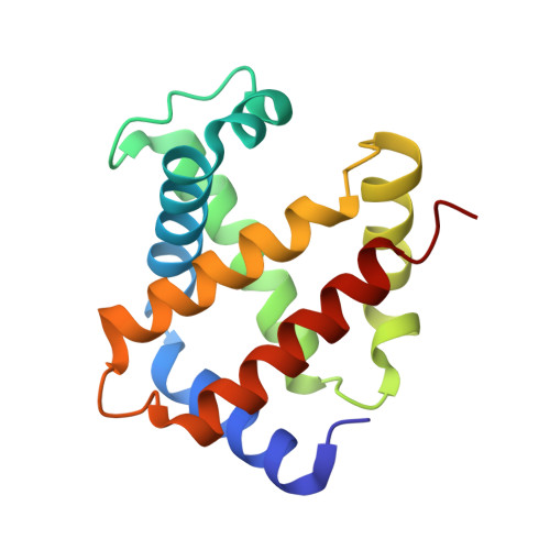
    The deer mouse, Peromyscus maniculatus, exhibits altitude-associated variation in hemoglobin oxygen affinity. To examine the structural basis of this functional variation, the structure of the hemoglobin was solved. Recombinant hemoglobin was expressed in Escherichia coli and was purified by ion-exchange chromatography. Recombinant hemoglobin was crystallized by the hanging-drop vapor-diffusion method using polyethylene glycol as a precipitant. The obtained orthorhombic crystal contained two subunits in the asymmetric unit. The refined structure was interpreted as the aquo-met form. Structural comparisons were performed among hemoglobins from deer mouse, house mouse and human. In contrast to human hemoglobin, deer mouse hemoglobin lacks the hydrogen bond between α1Trp14 in the A helix and α1Thr67 in the E helix owing to the Thr67Ala substitution. In addition, deer mouse hemoglobin has a unique hydrogen bond at the α1β1 interface between residues α1Cys34 and β1Ser128.


  • Organizational Affiliation

    School of Biological Sciences, University of Nebraska-Lincoln, Lincoln, Nebraska, USA.


Macromolecules
Find similar proteins by:  (by identity cutoff)  |  3D Structure
Entity ID: 1
MoleculeChains Sequence LengthOrganismDetailsImage
Alpha-globin141Peromyscus maniculatusMutation(s): 0 
Gene Names: HBA
UniProt
Find proteins for A4ZQ95 (Peromyscus maniculatus)
Explore A4ZQ95 
Go to UniProtKB:  A4ZQ95
Entity Groups  
Sequence Clusters30% Identity50% Identity70% Identity90% Identity95% Identity100% Identity
UniProt GroupA4ZQ95
Sequence Annotations
Expand
  • Reference Sequence
Find similar proteins by:  (by identity cutoff)  |  3D Structure
Entity ID: 2
MoleculeChains Sequence LengthOrganismDetailsImage
Beta globin146Peromyscus maniculatusMutation(s): 0 
Gene Names: HBB-T1HBB-T2
UniProt
Find proteins for C5MQT7 (Peromyscus maniculatus)
Explore C5MQT7 
Go to UniProtKB:  C5MQT7
Entity Groups  
Sequence Clusters30% Identity50% Identity70% Identity90% Identity95% Identity100% Identity
UniProt GroupC5MQT7
Sequence Annotations
Expand
  • Reference Sequence
Small Molecules
Ligands 1 Unique
IDChains Name / Formula / InChI Key2D Diagram3D Interactions
HEM
Query on HEM

Download Ideal Coordinates CCD File 
C [auth A],
D [auth B]
PROTOPORPHYRIN IX CONTAINING FE
C34 H32 Fe N4 O4
KABFMIBPWCXCRK-RGGAHWMASA-L
Experimental Data & Validation

Experimental Data

  • Method: X-RAY DIFFRACTION
  • Resolution: 1.78 Å
  • R-Value Free: 0.201 
  • R-Value Work: 0.162 
  • R-Value Observed: 0.165 
  • Space Group: C 2 2 21
Unit Cell:
Length ( Å )Angle ( ˚ )
a = 76.675α = 90
b = 80.061β = 90
c = 86.848γ = 90
Software Package:
Software NamePurpose
CrystalCleardata collection
PHENIXmodel building
PHENIXrefinement
HKL-2000data reduction
HKL-2000data scaling
PHENIXphasing

Structure Validation

View Full Validation Report


Ligand Structure Quality Assessment 


Entry History 

Deposition Data

Revision History  (Full details and data files)

  • Version 1.0: 2013-04-10
    Type: Initial release
  • Version 1.1: 2013-06-19
    Changes: Database references
  • Version 1.2: 2024-02-28
    Changes: Data collection, Database references, Derived calculations