4GZW

N2 neuraminidase D151G mutant of A/Tanzania/205/2010 H3N2 in complex with avian sialic acid receptor


Experimental Data Snapshot

  • Method: X-RAY DIFFRACTION
  • Resolution: 2.45 Å
  • R-Value Free: 0.246 
  • R-Value Work: 0.197 
  • R-Value Observed: 0.199 

wwPDB Validation   3D Report Full Report


Ligand Structure Quality Assessment 


This is version 2.1 of the entry. See complete history


Literature

Influenza virus neuraminidases with reduced enzymatic activity that avidly bind sialic Acid receptors.

Zhu, X.McBride, R.Nycholat, C.M.Yu, W.Paulson, J.C.Wilson, I.A.

(2012) J Virol 86: 13371-13383

  • DOI: https://doi.org/10.1128/JVI.01426-12
  • Primary Citation of Related Structures:  
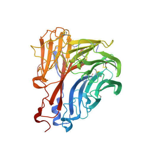
    4GZO, 4GZP, 4GZQ, 4GZS, 4GZT, 4GZW, 4GZX

  • PubMed Abstract: 

    Influenza virus neuraminidase (NA) cleaves off sialic acid from cellular receptors of hemagglutinin (HA) to enable progeny escape from infected cells. However, NA variants (D151G) of recent human H3N2 viruses have also been reported to bind receptors on red blood cells, but the nature of these receptors and the effect of the mutation on NA activity were not established. Here, we compare the functional and structural properties of a human H3N2 NA from A/Tanzania/205/2010 and its D151G mutant, which supports HA-independent receptor binding. While the wild-type NA efficiently cleaves sialic acid from both α2-6- and α2-3-linked glycans, the mutant exhibits much reduced enzymatic activity toward both types of sialosides. Conversely, while wild-type NA shows no detectable binding to sialosides, the D151G NA exhibits avid binding with broad specificity toward α2-3 sialosides. D151G NA binds the 3' sialyllactosamine (3'-SLN) and 6'-SLN sialosides with equilibrium dissociation constant (K(D)) values of 30.0 μM and 645 μM, respectively, which correspond to much higher affinities than the corresponding affinities (low mM) of HA to these glycans. Crystal structures of wild-type and mutant NAs reveal the structural basis for glycan binding in the active site by exclusively impairing the glycosidic bond hydrolysis step. The general significance of D151 among influenza virus NAs was further explored by introducing the D151G mutation into three N1 NAs and one N2 NA, which all exhibited reduced enzymatic activity and preferential binding to α2-3 sialosides. Since the enzymatic and binding activities of NAs are not routinely assessed, the potential for NA receptor binding to contribute to influenza virus biology may be underappreciated.


  • Organizational Affiliation

    Department of Molecular Biology, The Scripps Research Institute, La Jolla, California, USA.


Macromolecules
Find similar proteins by:  (by identity cutoff)  |  3D Structure
Entity ID: 1
MoleculeChains Sequence LengthOrganismDetailsImage
neuraminidase
A, B, C, D
393H3N2 subtypeMutation(s): 0 
UniProt
Find proteins for K7N5N3 (H3N2 subtype)
Explore K7N5N3 
Go to UniProtKB:  K7N5N3
Entity Groups  
Sequence Clusters30% Identity50% Identity70% Identity90% Identity95% Identity100% Identity
UniProt GroupK7N5N3
Sequence Annotations
Expand
  • Reference Sequence
Oligosaccharides

Help

Entity ID: 2
MoleculeChains Length2D Diagram Glycosylation3D Interactions
N-acetyl-alpha-neuraminic acid-(2-3)-beta-D-galactopyranose-(1-4)-2-acetamido-2-deoxy-beta-D-glucopyranose
E
3N/A
Glycosylation Resources
GlyTouCan:  G00065MO
GlyCosmos:  G00065MO
GlyGen:  G00065MO
Entity ID: 3
MoleculeChains Length2D Diagram Glycosylation3D Interactions
alpha-D-mannopyranose-(1-3)-[alpha-D-mannopyranose-(1-6)]beta-D-mannopyranose-(1-4)-2-acetamido-2-deoxy-beta-D-glucopyranose-(1-4)-2-acetamido-2-deoxy-beta-D-glucopyranose
F, J, L
5N-Glycosylation
Glycosylation Resources
GlyTouCan:  G22768VO
GlyCosmos:  G22768VO
GlyGen:  G22768VO
Entity ID: 4
MoleculeChains Length2D Diagram Glycosylation3D Interactions
N-acetyl-alpha-neuraminic acid-(2-3)-beta-D-galactopyranose-(1-3)-2-acetamido-2-deoxy-beta-D-glucopyranose
G
3N/A
Glycosylation Resources
GlyTouCan:  G46776WR
GlyCosmos:  G46776WR
GlyGen:  G46776WR
Entity ID: 5
MoleculeChains Length2D Diagram Glycosylation3D Interactions
alpha-D-mannopyranose-(1-3)-beta-D-mannopyranose-(1-4)-2-acetamido-2-deoxy-beta-D-glucopyranose-(1-4)-2-acetamido-2-deoxy-beta-D-glucopyranose
H
4N-Glycosylation
Glycosylation Resources
GlyTouCan:  G81315DD
GlyCosmos:  G81315DD
GlyGen:  G81315DD
Entity ID: 6
MoleculeChains Length2D Diagram Glycosylation3D Interactions
N-acetyl-alpha-neuraminic acid-(2-3)-beta-D-galactopyranose
I
2N/A
Glycosylation Resources
GlyTouCan:  G30207PZ
GlyCosmos:  G30207PZ
GlyGen:  G30207PZ
Entity ID: 7
MoleculeChains Length2D Diagram Glycosylation3D Interactions
2-acetamido-2-deoxy-beta-D-glucopyranose-(1-4)-2-acetamido-2-deoxy-beta-D-glucopyranose
K
2N-Glycosylation
Glycosylation Resources
GlyTouCan:  G42666HT
GlyCosmos:  G42666HT
GlyGen:  G42666HT
Entity ID: 8
MoleculeChains Length2D Diagram Glycosylation3D Interactions
2-acetamido-2-deoxy-beta-D-glucopyranose-(1-4)-[alpha-L-fucopyranose-(1-6)]2-acetamido-2-deoxy-beta-D-glucopyranose
M
3N-Glycosylation
Glycosylation Resources
GlyTouCan:  G21290RB
GlyCosmos:  G21290RB
GlyGen:  G21290RB
Small Molecules
Ligands 3 Unique
IDChains Name / Formula / InChI Key2D Diagram3D Interactions
SIA
Query on SIA

Download Ideal Coordinates CCD File 
BA [auth D]N-acetyl-alpha-neuraminic acid
C11 H19 N O9
SQVRNKJHWKZAKO-YRMXFSIDSA-N
NAG
Query on NAG

Download Ideal Coordinates CCD File 
CA [auth D]
DA [auth D]
N [auth A]
O [auth A]
P [auth A]
CA [auth D],
DA [auth D],
N [auth A],
O [auth A],
P [auth A],
Q [auth A],
S [auth B],
T [auth B],
U [auth B],
V [auth B],
X [auth C],
Y [auth C]
2-acetamido-2-deoxy-beta-D-glucopyranose
C8 H15 N O6
OVRNDRQMDRJTHS-FMDGEEDCSA-N
CA
Query on CA

Download Ideal Coordinates CCD File 
AA [auth D],
EA [auth D],
R [auth A],
W [auth B],
Z [auth C]
CALCIUM ION
Ca
BHPQYMZQTOCNFJ-UHFFFAOYSA-N
Binding Affinity Annotations 
IDSourceBinding Affinity
SIA PDBBind:  4GZW Kd: 3.00e+4 (nM) from 1 assay(s)
Biologically Interesting Molecules (External Reference) 1 Unique
Experimental Data & Validation

Experimental Data

  • Method: X-RAY DIFFRACTION
  • Resolution: 2.45 Å
  • R-Value Free: 0.246 
  • R-Value Work: 0.197 
  • R-Value Observed: 0.199 
  • Space Group: P 1 21 1
Unit Cell:
Length ( Å )Angle ( ˚ )
a = 82.439α = 90
b = 110.555β = 97.79
c = 109.928γ = 90
Software Package:
Software NamePurpose
HKL-2000data collection
PHASERphasing
REFMACrefinement
HKL-2000data reduction
HKL-2000data scaling

Structure Validation

View Full Validation Report


Ligand Structure Quality Assessment 


Entry History 

Deposition Data

Revision History  (Full details and data files)

  • Version 1.0: 2012-10-17
    Type: Initial release
  • Version 1.1: 2012-12-05
    Changes: Database references
  • Version 2.0: 2020-07-29
    Type: Remediation
    Reason: Carbohydrate remediation
    Changes: Advisory, Atomic model, Data collection, Derived calculations, Structure summary
  • Version 2.1: 2023-09-13
    Changes: Data collection, Database references, Refinement description, Structure summary