4GXX

Crystal structure of the "avianized" 1918 influenza virus hemagglutinin


Experimental Data Snapshot

  • Method: X-RAY DIFFRACTION
  • Resolution: 1.80 Å
  • R-Value Free: 0.209 
  • R-Value Work: 0.180 
  • R-Value Observed: 0.182 

wwPDB Validation   3D Report Full Report


This is version 2.1 of the entry. See complete history


Literature

Influenza Human Monoclonal Antibody 1F1 Interacts with Three Major Antigenic Sites and Residues Mediating Human Receptor Specificity in H1N1 Viruses.

Tsibane, T.Ekiert, D.C.Krause, J.C.Martinez, O.Crowe, J.E.Wilson, I.A.Basler, C.F.

(2012) PLoS Pathog 8: e1003067-e1003067

  • DOI: https://doi.org/10.1371/journal.ppat.1003067
  • Primary Citation of Related Structures:  
    4GXU, 4GXV, 4GXX

  • PubMed Abstract: 

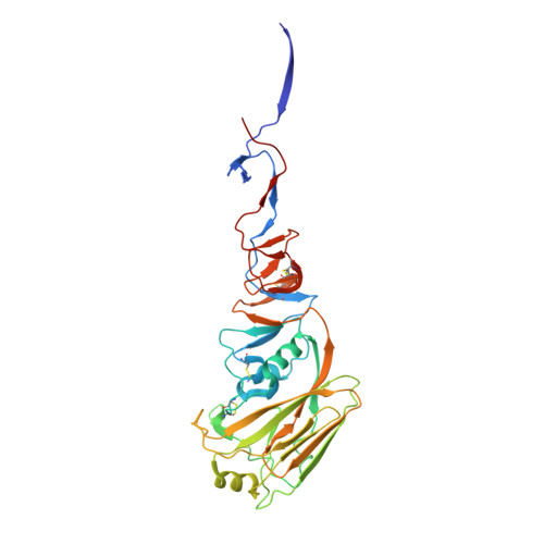
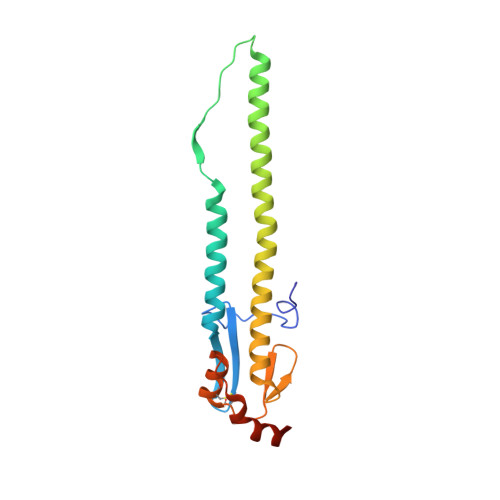
    Most monoclonal antibodies (mAbs) to the influenza A virus hemagglutinin (HA) head domain exhibit very limited breadth of inhibitory activity due to antigenic drift in field strains. However, mAb 1F1, isolated from a 1918 influenza pandemic survivor, inhibits select human H1 viruses (1918, 1943, 1947, and 1977 isolates). The crystal structure of 1F1 in complex with the 1918 HA shows that 1F1 contacts residues that are classically defined as belonging to three distinct antigenic sites, Sa, Sb and Ca(2). The 1F1 heavy chain also reaches into the receptor binding site (RBS) and interacts with residues that contact sialoglycan receptors and determine HA receptor specificity. The 1F1 epitope is remarkably similar to the previously described murine HC63 H3 epitope, despite significant sequence differences between H1 and H3 HAs. Both antibodies potently inhibit receptor binding, but only HC63 can block the pH-induced conformational changes in HA that drive membrane fusion. Contacts within the RBS suggested that 1F1 may be sensitive to changes that alter HA receptor binding activity. Affinity assays confirmed that sequence changes that switch the HA to avian receptor specificity affect binding of 1F1 and a mAb possessing a closely related heavy chain, 1I20. To characterize 1F1 cross-reactivity, additional escape mutant selection and site-directed mutagenesis were performed. Residues 190 and 227 in the 1F1 epitope were found to be critical for 1F1 reactivity towards 1918, 1943 and 1977 HAs, as well as for 1I20 reactivity towards the 1918 HA. Therefore, 1F1 heavy-chain interactions with conserved RBS residues likely contribute to its ability to inhibit divergent HAs.


  • Organizational Affiliation

    Department of Microbiology, Mount Sinai School of Medicine, New York City, New York, United States of America.


Macromolecules
Find similar proteins by:  (by identity cutoff)  |  3D Structure
Entity ID: 1
MoleculeChains Sequence LengthOrganismDetailsImage
Hemagglutinin HA1 chain
A, C, E
331Influenza A virus (A/Brevig Mission/1/1918(H1N1))Mutation(s): 2 
Gene Names: HA
UniProt
Find proteins for Q9WFX3 (Influenza A virus (strain A/Brevig Mission/1/1918 H1N1))
Explore Q9WFX3 
Go to UniProtKB:  Q9WFX3
Entity Groups  
Sequence Clusters30% Identity50% Identity70% Identity90% Identity95% Identity100% Identity
UniProt GroupQ9WFX3
Sequence Annotations
Expand
  • Reference Sequence
Find similar proteins by:  (by identity cutoff)  |  3D Structure
Entity ID: 2
MoleculeChains Sequence LengthOrganismDetailsImage
Hemagglutinin HA2 chain
B, D, F
176Influenza A virus (A/Brevig Mission/1/1918(H1N1))Mutation(s): 0 
Gene Names: HA
UniProt
Find proteins for Q9WFX3 (Influenza A virus (strain A/Brevig Mission/1/1918 H1N1))
Explore Q9WFX3 
Go to UniProtKB:  Q9WFX3
Entity Groups  
Sequence Clusters30% Identity50% Identity70% Identity90% Identity95% Identity100% Identity
UniProt GroupQ9WFX3
Sequence Annotations
Expand
  • Reference Sequence
Oligosaccharides

Help

Entity ID: 3
MoleculeChains Length2D Diagram Glycosylation3D Interactions
alpha-D-mannopyranose-(1-3)-beta-D-mannopyranose-(1-4)-2-acetamido-2-deoxy-beta-D-glucopyranose-(1-4)-2-acetamido-2-deoxy-beta-D-glucopyranose
G
4N-Glycosylation
Glycosylation Resources
GlyTouCan:  G81315DD
GlyCosmos:  G81315DD
GlyGen:  G81315DD
Entity ID: 4
MoleculeChains Length2D Diagram Glycosylation3D Interactions
alpha-D-mannopyranose-(1-3)-[alpha-D-mannopyranose-(1-6)]beta-D-mannopyranose-(1-4)-2-acetamido-2-deoxy-beta-D-glucopyranose-(1-4)-2-acetamido-2-deoxy-beta-D-glucopyranose
H
5N-Glycosylation
Glycosylation Resources
GlyTouCan:  G22768VO
GlyCosmos:  G22768VO
GlyGen:  G22768VO
Entity ID: 5
MoleculeChains Length2D Diagram Glycosylation3D Interactions
beta-D-mannopyranose-(1-4)-2-acetamido-2-deoxy-beta-D-glucopyranose-(1-4)-2-acetamido-2-deoxy-beta-D-glucopyranose
I
3N-Glycosylation
Glycosylation Resources
GlyTouCan:  G15407YE
GlyCosmos:  G15407YE
GlyGen:  G15407YE
Experimental Data & Validation

Experimental Data

  • Method: X-RAY DIFFRACTION
  • Resolution: 1.80 Å
  • R-Value Free: 0.209 
  • R-Value Work: 0.180 
  • R-Value Observed: 0.182 
  • Space Group: P 1 21 1
Unit Cell:
Length ( Å )Angle ( ˚ )
a = 71.79α = 90
b = 241.51β = 119.77
c = 72.03γ = 90
Software Package:
Software NamePurpose
Blu-Icedata collection
PHASERphasing
PHENIXrefinement
HKL-2000data reduction
HKL-2000data scaling

Structure Validation

View Full Validation Report


Entry History 

Deposition Data

Revision History  (Full details and data files)

  • Version 1.0: 2012-12-19
    Type: Initial release
  • Version 1.1: 2012-12-26
    Changes: Database references, Derived calculations
  • Version 2.0: 2020-07-29
    Type: Remediation
    Reason: Carbohydrate remediation
    Changes: Atomic model, Data collection, Database references, Derived calculations, Structure summary
  • Version 2.1: 2023-09-13
    Changes: Data collection, Database references, Refinement description, Structure summary