4GWJ

His 62 mutant of the lectin binding domain of Lectinolysin complexed with Lewis b


Experimental Data Snapshot

  • Method: X-RAY DIFFRACTION
  • Resolution: 1.60 Å
  • R-Value Free: 0.210 
  • R-Value Work: 0.179 
  • R-Value Observed: 0.181 

wwPDB Validation   3D Report Full Report


This is version 2.1 of the entry. See complete history


Literature

Manipulating the Lewis antigen specificity of the cholesterol-dependent cytolysin lectinolysin

Lawrence, S.L.Feil, S.C.Holien, J.K.Kuiper, M.J.Doughty, L.Dolezal, O.Mulhern, T.D.Tweten, R.K.Parker, M.W.

(2012) Front Immunol 3: 330-330

  • DOI: https://doi.org/10.3389/fimmu.2012.00330
  • Primary Citation of Related Structures:  
    4GWI, 4GWJ

  • PubMed Abstract: 

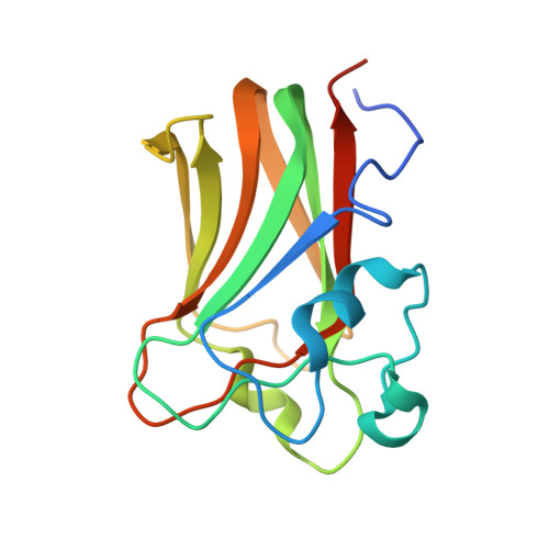
    The cholesterol-dependent cytolysins (CDCs) attack cells by punching large holes in their membranes. Lectinolysin from Streptococcus mitis is unique among CDCs due to the presence of an N-terminal lectin domain that enhances the pore-forming activity of the toxin. We recently determined the crystal structures of the lectin domain in complex with various glycans. These structures revealed the molecular basis for the Lewis antigen specificity of the toxin. Based on this information we have used in silico molecular modeling to design a mutant toxin, which we predicted would increase its specificity for Lewis y, an antigen found on the surface of cancer cells. Surprisingly, we found by surface plasmon resonance binding experiments that the resultant mutant lectin domain exhibited higher specificity for Lewis b antigens instead. We then undertook comparative crystallographic and molecular dynamics simulation studies of the wild-type and mutant lectin domains to understand the molecular basis for the disparity between the theoretical and experimental results. The crystallographic results revealed that the net number of interactions between Lewis y and wild-type versus mutant was unchanged whereas there was a loss of a hydrogen bond between mutant and Lewis b compared to wild-type. In contrast, the molecular dynamics studies revealed that the Lewis b antigen spent more time in the binding pocket of the mutant compared to wild-type and the reverse was true for Lewis y. The results of these simulation studies are consistent with the conclusions drawn from the surface plasmon resonance studies. This work is part of a program to engineer lectinolysin so that it will target and kill specific cells in human diseases.


  • Organizational Affiliation

    Biota Structural Biology Laboratory and Australian Cancer Research Foundation Rational Drug Discovery Centre, St. Vincent's Institute of Medical Research Fitzroy, VIC, Australia.


Macromolecules
Find similar proteins by:  (by identity cutoff)  |  3D Structure
Entity ID: 1
MoleculeChains Sequence LengthOrganismDetailsImage
Platelet aggregation factor Sm-hPAF153Streptococcus mitisMutation(s): 2 
Gene Names: samhpaf
UniProt
Find proteins for Q2PHL4 (Streptococcus mitis)
Explore Q2PHL4 
Go to UniProtKB:  Q2PHL4
Entity Groups  
Sequence Clusters30% Identity50% Identity70% Identity90% Identity95% Identity100% Identity
UniProt GroupQ2PHL4
Sequence Annotations
Expand
  • Reference Sequence
Oligosaccharides

Help

Entity ID: 2
MoleculeChains Length2D Diagram Glycosylation3D Interactions
alpha-L-fucopyranose-(1-2)-beta-D-galactopyranose-(1-3)-[alpha-L-fucopyranose-(1-4)]2-acetamido-2-deoxy-beta-D-glucopyranose
B
4N/A
Glycosylation Resources
GlyTouCan:  G00048MO
GlyCosmos:  G00048MO
GlyGen:  G00048MO
Biologically Interesting Molecules (External Reference) 1 Unique
Experimental Data & Validation

Experimental Data

  • Method: X-RAY DIFFRACTION
  • Resolution: 1.60 Å
  • R-Value Free: 0.210 
  • R-Value Work: 0.179 
  • R-Value Observed: 0.181 
  • Space Group: P 43 21 2
Unit Cell:
Length ( Å )Angle ( ˚ )
a = 66.923α = 90
b = 66.923β = 90
c = 99.387γ = 90
Software Package:
Software NamePurpose
DENZOdata reduction
SCALEPACKdata scaling
REFMACrefinement
PDB_EXTRACTdata extraction
HKL-2000data collection

Structure Validation

View Full Validation Report


Entry History 

Deposition Data

  • Released Date: 2012-11-21 
  • Deposition Author(s): Feil, S.C.

Revision History  (Full details and data files)

  • Version 1.0: 2012-11-21
    Type: Initial release
  • Version 1.1: 2014-03-12
    Changes: Database references
  • Version 1.2: 2017-11-15
    Changes: Advisory, Refinement description
  • Version 2.0: 2020-07-29
    Type: Remediation
    Reason: Carbohydrate remediation
    Changes: Atomic model, Data collection, Database references, Derived calculations, Structure summary
  • Version 2.1: 2023-11-08
    Changes: Advisory, Data collection, Database references, Refinement description, Structure summary