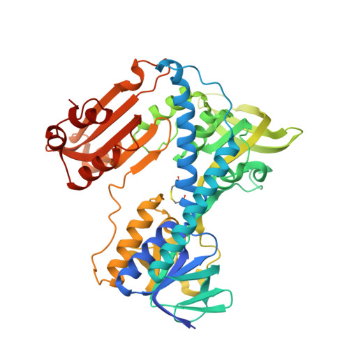
4GR1

THE BINDING OF THE RETRO-ANALOGUE OF GLUTATHIONE DISULFIDE TO GLUTATHIONE REDUCTASE


Experimental Data Snapshot

  • Method: X-RAY DIFFRACTION
  • Resolution: 2.40 Å
  • R-Value Observed: 0.151 

wwPDB Validation   3D Report Full Report


Ligand Structure Quality Assessment 


This is version 1.2 of the entry. See complete history


Literature

The binding of the retro-analogue of glutathione disulfide to glutathione reductase.

Janes, W.Schulz, G.E.

(1990) J Biol Chem 265: 10443-10445

  • DOI: https://doi.org/10.2210/pdb4gr1/pdb
  • Primary Citation of Related Structures:  
    4GR1

  • PubMed Abstract: 

    The retro-analogue of glutathione disulfide was bound to the GSSG binding site of crystalline glutathione reductase. The binding mode revealed why the analogue is a very poor substrate in enzyme catalysis. The observed binding mode difference between natural substrate and retro-analogue is explained.


  • Organizational Affiliation

    Institut für Organische Chemie und Biochemie, Universität Freiburg, Federal Republic of Germany.


Macromolecules
Find similar proteins by:  (by identity cutoff)  |  3D Structure
Entity ID: 1
MoleculeChains Sequence LengthOrganismDetailsImage
GLUTATHIONE REDUCTASE478Homo sapiensMutation(s): 0 
EC: 1.6.4.2
UniProt & NIH Common Fund Data Resources
Find proteins for P00390 (Homo sapiens)
Explore P00390 
Go to UniProtKB:  P00390
PHAROS:  P00390
GTEx:  ENSG00000104687 
Entity Groups  
Sequence Clusters30% Identity50% Identity70% Identity90% Identity95% Identity100% Identity
UniProt GroupP00390
Sequence Annotations
Expand
  • Reference Sequence
Small Molecules
Ligands 3 Unique
IDChains Name / Formula / InChI Key2D Diagram3D Interactions
FAD
Query on FAD

Download Ideal Coordinates CCD File 
C [auth A]FLAVIN-ADENINE DINUCLEOTIDE
C27 H33 N9 O15 P2
VWWQXMAJTJZDQX-UYBVJOGSSA-N
RGS
Query on RGS

Download Ideal Coordinates CCD File 
D [auth A]4N-MALONYL-CYSTEINYL-2,4-DIAMINOBUTYRATE DISULFIDE
C20 H32 N6 O12 S2
MRZMTLNHPCSZCE-NNYUYHANSA-N
PO4
Query on PO4

Download Ideal Coordinates CCD File 
B [auth A]PHOSPHATE ION
O4 P
NBIIXXVUZAFLBC-UHFFFAOYSA-K
Experimental Data & Validation

Experimental Data

  • Method: X-RAY DIFFRACTION
  • Resolution: 2.40 Å
  • R-Value Observed: 0.151 
  • Space Group: B 1 1 2
Unit Cell:
Length ( Å )Angle ( ˚ )
a = 119.8α = 90
b = 84.5β = 90
c = 63.2γ = 58.7
Software Package:
Software NamePurpose
TNTrefinement

Structure Validation

View Full Validation Report


Ligand Structure Quality Assessment 


Entry History 

Deposition Data

Revision History  (Full details and data files)

  • Version 1.0: 1991-10-15
    Type: Initial release
  • Version 1.1: 2008-03-03
    Changes: Version format compliance
  • Version 1.2: 2011-07-13
    Changes: Derived calculations, Version format compliance