


Crystal Structure of the Substrate-Free and the Product-Bound forms of HmuO, a Heme Oxygenase from Corynebacterium diphtheriae
Unno, M., Ardevol, A., Rovira, C., Ikeda-Saito, M.To be published.
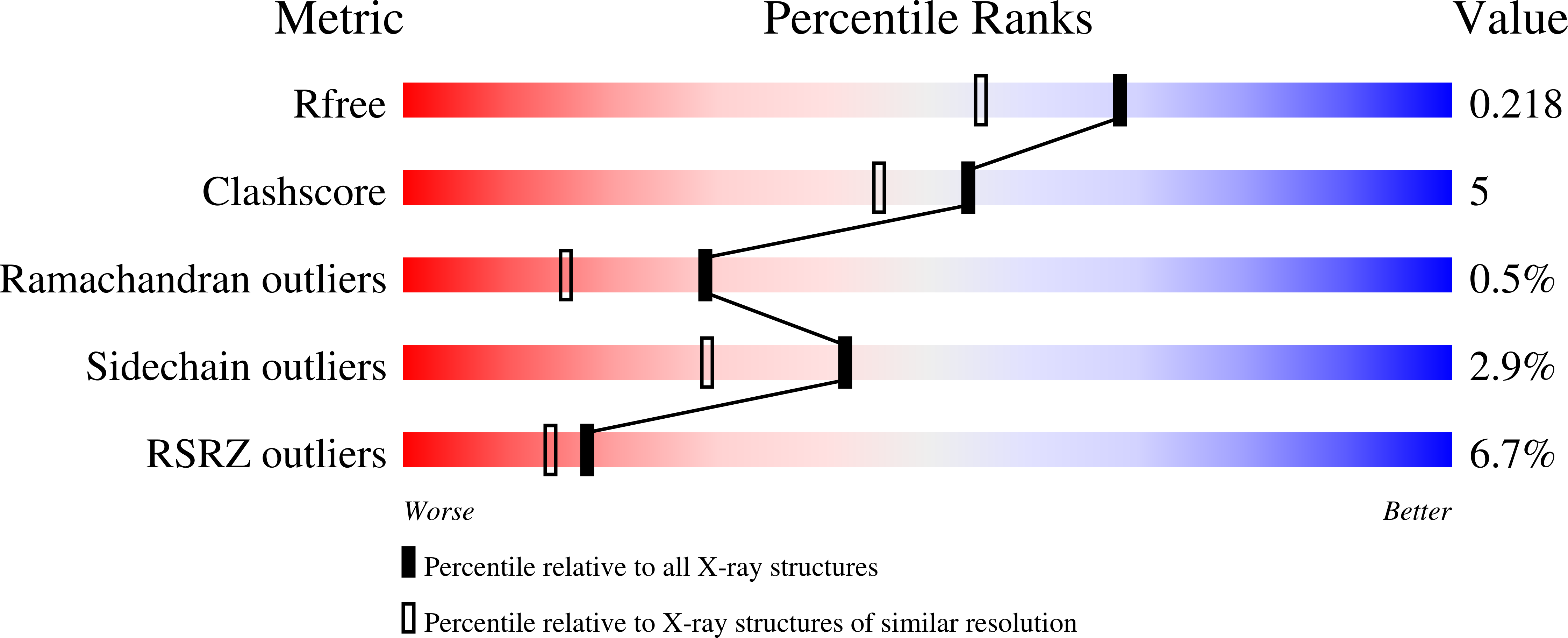
Experimental Data Snapshot
wwPDB Validation 3D Report Full Report
Entity ID: 1 | |||||
|---|---|---|---|---|---|
| Molecule | Chains | Sequence Length | Organism | Details | Image |
| Heme oxygenase | 215 | Corynebacterium diphtheriae | Mutation(s): 0 EC: 1.14.99.3 | 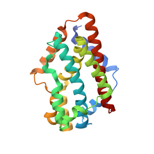 | |
UniProt | |||||
Find proteins for Q54AI1 (Corynebacterium diphtheriae) Explore Q54AI1 Go to UniProtKB: Q54AI1 | |||||
Entity Groups | |||||
| Sequence Clusters | 30% Identity50% Identity70% Identity90% Identity95% Identity100% Identity | ||||
| UniProt Group | Q54AI1 | ||||
Sequence AnnotationsExpand | |||||
| |||||
| Ligands 2 Unique | |||||
|---|---|---|---|---|---|
| ID | Chains | Name / Formula / InChI Key | 2D Diagram | 3D Interactions | |
| GOL Query on GOL | B [auth A] C [auth A] D [auth A] E [auth A] F [auth A] | GLYCEROL C3 H8 O3 PEDCQBHIVMGVHV-UHFFFAOYSA-N |  | ||
| NA Query on NA | H [auth A], I [auth A] | SODIUM ION Na FKNQFGJONOIPTF-UHFFFAOYSA-N |  | ||
| Length ( Å ) | Angle ( ˚ ) |
|---|---|
| a = 57.489 | α = 90 |
| b = 61.916 | β = 90 |
| c = 63.086 | γ = 90 |
| Software Name | Purpose |
|---|---|
| ADSC | data collection |
| CNS | refinement |
| REFMAC | refinement |
| HKL-2000 | data reduction |
| HKL-2000 | data scaling |
| CNS | phasing |
RCSB PDB (citation) is hosted by
RCSB PDB is a member of the

