4GLR

Structure of the anti-ptau Fab (pT231/pS235_1) in complex with phosphoepitope pT231/pS235


Experimental Data Snapshot

  • Method: X-RAY DIFFRACTION
  • Resolution: 1.90 Å
  • R-Value Free: 0.208 
  • R-Value Work: 0.179 
  • R-Value Observed: 0.181 

wwPDB Validation   3D Report Full Report


This is version 1.4 of the entry. See complete history


Literature

An Ultra-specific Avian Antibody to Phosphorylated Tau Protein Reveals a Unique Mechanism for Phosphoepitope Recognition.

Shih, H.H.Tu, C.Cao, W.Klein, A.Ramsey, R.Fennell, B.J.Lambert, M.Ni Shuilleabhain, D.Autin, B.Kouranova, E.Laxmanan, S.Braithwaite, S.Wu, L.Ait-Zahra, M.Milici, A.J.Dumin, J.A.Lavallie, E.R.Arai, M.Corcoran, C.Paulsen, J.E.Gill, D.Cunningham, O.Bard, J.Mosyak, L.Finlay, W.J.

(2012) J Biol Chem 287: 44425-44434

  • DOI: https://doi.org/10.1074/jbc.M112.415935
  • Primary Citation of Related Structures:  
    4GLR

  • PubMed Abstract: 

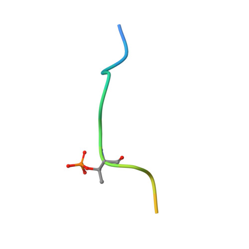
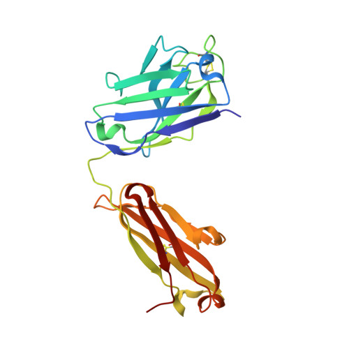
    Highly specific antibodies to phosphoepitopes are valuable tools to study phosphorylation in disease states, but their discovery is largely empirical, and the molecular mechanisms mediating phosphospecific binding are poorly understood. Here, we report the generation and characterization of extremely specific recombinant chicken antibodies to three phosphoepitopes on the Alzheimer disease-associated protein tau. Each antibody shows full specificity for a single phosphopeptide. The chimeric IgG pT231/pS235_1 exhibits a K(D) of 0.35 nm in 1:1 binding to its cognate phosphopeptide. This IgG is murine ortholog-cross-reactive, specifically recognizing the pathological form of tau in brain samples from Alzheimer patients and a mouse model of tauopathy. To better understand the underlying binding mechanisms allowing such remarkable specificity, we determined the structure of pT231/pS235_1 Fab in complex with its cognate phosphopeptide at 1.9 Å resolution. The Fab fragment exhibits novel complementarity determining region (CDR) structures with a "bowl-like" conformation in CDR-H2 that tightly and specifically interacts with the phospho-Thr-231 phosphate group, as well as a long, disulfide-constrained CDR-H3 that mediates peptide recognition. This binding mechanism differs distinctly from either peptide- or hapten-specific antibodies described to date. Surface plasmon resonance analyses showed that pT231/pS235_1 binds a truly compound epitope, as neither phosphorylated Ser-235 nor free peptide shows any measurable binding affinity.


  • Organizational Affiliation

    Global Biotherapeutics Technologies, Pfizer Global Research & Development, Cambridge, Massachusetts 02140, USA.


Macromolecules

Find similar proteins by:  Sequence   |   3D Structure  

Entity ID: 1
MoleculeChains Sequence LengthOrganismDetailsImage
phospho-peptide
A, B
18Homo sapiensMutation(s): 0 
UniProt & NIH Common Fund Data Resources
Find proteins for P10636 (Homo sapiens)
Explore P10636 
Go to UniProtKB:  P10636
PHAROS:  P10636
GTEx:  ENSG00000186868 
Entity Groups  
Sequence Clusters30% Identity50% Identity70% Identity90% Identity95% Identity100% Identity
UniProt GroupP10636
Sequence Annotations
Expand
  • Reference Sequence
Find similar proteins by:  (by identity cutoff)  |  3D Structure
Entity ID: 2
MoleculeChains Sequence LengthOrganismDetailsImage
anti-ptau heavy chainC [auth H],
E [auth J]
229Gallus gallusMutation(s): 0 
Entity Groups  
Sequence Clusters30% Identity50% Identity70% Identity90% Identity95% Identity100% Identity
Sequence Annotations
Expand
  • Reference Sequence
Find similar proteins by:  (by identity cutoff)  |  3D Structure
Entity ID: 3
MoleculeChains Sequence LengthOrganismDetailsImage
anti-ptau light chainD [auth I],
F [auth K]
212Gallus gallusMutation(s): 0 
Entity Groups  
Sequence Clusters30% Identity50% Identity70% Identity90% Identity95% Identity100% Identity
Sequence Annotations
Expand
  • Reference Sequence
Small Molecules
Modified Residues  2 Unique
IDChains TypeFormula2D DiagramParent
SEP
Query on SEP
A, B
L-PEPTIDE LINKINGC3 H8 N O6 PSER
TPO
Query on TPO
A, B
L-PEPTIDE LINKINGC4 H10 N O6 PTHR
Experimental Data & Validation

Experimental Data

  • Method: X-RAY DIFFRACTION
  • Resolution: 1.90 Å
  • R-Value Free: 0.208 
  • R-Value Work: 0.179 
  • R-Value Observed: 0.181 
  • Space Group: P 21 21 2
Unit Cell:
Length ( Å )Angle ( ˚ )
a = 68.5α = 90
b = 216.5β = 90
c = 70γ = 90
Software Package:
Software NamePurpose
HKL-2000data collection
PHASERphasing
BUSTERrefinement
PROCESSdata reduction
SCALAdata scaling

Structure Validation

View Full Validation Report


Entry History 

Deposition Data

Revision History  (Full details and data files)

  • Version 1.0: 2012-11-21
    Type: Initial release
  • Version 1.1: 2013-01-02
    Changes: Database references
  • Version 1.2: 2013-01-16
    Changes: Database references
  • Version 1.3: 2013-10-02
    Changes: Source and taxonomy
  • Version 1.4: 2023-09-13
    Changes: Data collection, Database references, Derived calculations, Refinement description