4GGN

Malaria invasion machinery protein complex


Experimental Data Snapshot

  • Method: X-RAY DIFFRACTION
  • Resolution: 2.29 Å
  • R-Value Free: 0.279 
  • R-Value Work: 0.226 
  • R-Value Observed: 0.228 

wwPDB Validation   3D Report Full Report


This is version 1.3 of the entry. See complete history


Literature

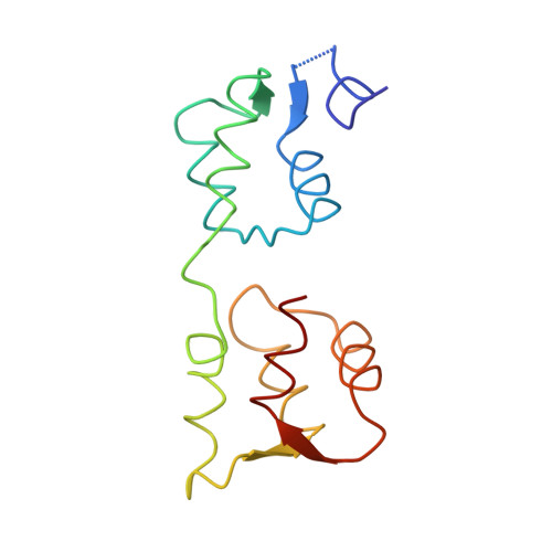
The structure of the D3 domain of Plasmodium falciparum myosin tail interacting protein MTIP in complex with a nanobody.

Khamrui, S.Turley, S.Pardon, E.Steyaert, J.Fan, E.Verlinde, C.L.Bergman, L.W.Hol, W.G.

(2013) Mol Biochem Parasitol 190: 87-91

  • DOI: https://doi.org/10.1016/j.molbiopara.2013.06.003
  • Primary Citation of Related Structures:  
    4GFT, 4GGN

  • PubMed Abstract: 

    Apicomplexan parasites enter host cells by many sophisticated steps including use of an ATP-powered invasion machinery. The machinery consists of multiple proteins, including a special myosin (MyoA) which moves along an actin fiber and which is connected to the myosin tail interaction protein (MTIP). Here we report a crystal structure of the major MyoA-binding domain (D3) of Plasmodium falciparum MTIP in complex with an anti-MTIP nanobody. In this complex, the MyoA-binding groove in MTIP-D3 is considerably less accessible than when occupied by the MyoA helix, due to a shift of two helices. The nanobody binds to an area slightly overlapping with the MyoA binding groove, covering a hydrophobic region next to the groove entrance. This provides a new avenue for arriving at compounds interfering with the invasion machinery since small molecules binding simultaneously to the nanobody binding site and the adjacent MyoA binding groove would prevent MyoA binding by MTIP.


  • Organizational Affiliation

    Department of Biochemistry, Biomolecular Structure Center, School of Medicine, University of Washington, Seattle, WA 98195, United States.


Macromolecules
Find similar proteins by:  (by identity cutoff)  |  3D Structure
Entity ID: 1
MoleculeChains Sequence LengthOrganismDetailsImage
Myosin A tail domain interacting protein MTIP
A, B, C
127Plasmodium knowlesi strain HMutation(s): 0 
Gene Names: PKH_146380
UniProt
Find proteins for A0A384KCF1 (Plasmodium knowlesi (strain H))
Explore A0A384KCF1 
Go to UniProtKB:  A0A384KCF1
Entity Groups  
Sequence Clusters30% Identity50% Identity70% Identity90% Identity95% Identity100% Identity
UniProt GroupA0A384KCF1
Sequence Annotations
Expand
  • Reference Sequence

Find similar proteins by:  Sequence   |   3D Structure  

Entity ID: 2
MoleculeChains Sequence LengthOrganismDetailsImage
Myosin-A
D, E, F
15Plasmodium yoelii yoeliiMutation(s): 0 
Gene Names: PY01232
UniProt
Find proteins for Q7RQ71 (Plasmodium yoelii yoelii)
Explore Q7RQ71 
Go to UniProtKB:  Q7RQ71
Entity Groups  
Sequence Clusters30% Identity50% Identity70% Identity90% Identity95% Identity100% Identity
UniProt GroupQ7RQ71
Sequence Annotations
Expand
  • Reference Sequence
Experimental Data & Validation

Experimental Data

  • Method: X-RAY DIFFRACTION
  • Resolution: 2.29 Å
  • R-Value Free: 0.279 
  • R-Value Work: 0.226 
  • R-Value Observed: 0.228 
  • Space Group: P 21 21 21
Unit Cell:
Length ( Å )Angle ( ˚ )
a = 82.56α = 90
b = 83.403β = 90
c = 84.316γ = 90
Software Package:
Software NamePurpose
HKL-2000data collection
PHASERphasing
REFMACrefinement
HKL-2000data reduction
HKL-2000data scaling

Structure Validation

View Full Validation Report


Entry History 

Deposition Data

Revision History  (Full details and data files)

  • Version 1.0: 2013-07-03
    Type: Initial release
  • Version 1.1: 2013-08-07
    Changes: Atomic model
  • Version 1.2: 2015-12-09
    Changes: Database references
  • Version 1.3: 2024-02-28
    Changes: Data collection, Database references