4GDE

Crystal structure of NADPH-reduced Aspergillus fumigatus UDP-galactopyranose


Experimental Data Snapshot

  • Method: X-RAY DIFFRACTION
  • Resolution: 2.20 Å
  • R-Value Free: 0.227 
  • R-Value Work: 0.201 
  • R-Value Observed: 0.202 

wwPDB Validation   3D Report Full Report


Ligand Structure Quality Assessment 


This is version 1.3 of the entry. See complete history


Literature

Identification of the NAD(P)H Binding Site of Eukaryotic UDP-Galactopyranose Mutase.

Dhatwalia, R.Singh, H.Solano, L.M.Oppenheimer, M.Robinson, R.M.Ellerbrock, J.F.Sobrado, P.Tanner, J.J.

(2012) J Am Chem Soc 134: 18132-18138

  • DOI: https://doi.org/10.1021/ja308188z
  • Primary Citation of Related Structures:  
    4GDE, 5VWT, 5VWU

  • PubMed Abstract: 

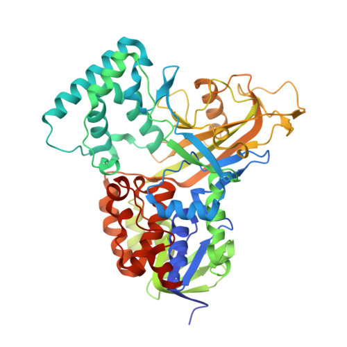
    UDP-galactopyranose mutase (UGM) plays an essential role in galactofuranose biosynthesis in microorganisms by catalyzing the conversion of UDP-galactopyranose to UDP-galactofuranose. The enzyme has gained attention recently as a promising target for the design of new antifungal, antitrypanosomal, and antileishmanial agents. Here we report the first crystal structure of UGM complexed with its redox partner NAD(P)H. Kinetic protein crystallography was used to obtain structures of oxidized Aspergillus fumigatus UGM (AfUGM) complexed with NADPH and NADH, as well as reduced AfUGM after dissociation of NADP(+). NAD(P)H binds with the nicotinamide near the FAD isoalloxazine and the ADP moiety extending toward the mobile 200s active site flap. The nicotinamide riboside binding site overlaps that of the substrate galactopyranose moiety, and thus NADPH and substrate binding are mutually exclusive. On the other hand, the pockets for the adenine of NADPH and uracil of the substrate are distinct and separated by only 6 Å, which raises the possibility of designing novel inhibitors that bind both sites. All 12 residues that contact NADP(H) are conserved among eukaryotic UGMs. Residues that form the AMP pocket are absent in bacterial UGMs, which suggests that eukaryotic and bacterial UGMs have different NADP(H) binding sites. The structures address the longstanding question of how UGM binds NAD(P)H and provide new opportunities for drug discovery.


  • Organizational Affiliation

    Department of Chemistry, University of Missouri-Columbia, Columbia, Missouri 65211, USA.


Macromolecules
Find similar proteins by:  (by identity cutoff)  |  3D Structure
Entity ID: 1
MoleculeChains Sequence LengthOrganismDetailsImage
UDP-galactopyranose mutase
A, B, C, D
513Aspergillus fumigatusMutation(s): 2 
Gene Names: GLF
EC: 5.4.99.9
UniProt
Find proteins for Q4W1X2 (Aspergillus fumigatus)
Explore Q4W1X2 
Go to UniProtKB:  Q4W1X2
Entity Groups  
Sequence Clusters30% Identity50% Identity70% Identity90% Identity95% Identity100% Identity
UniProt GroupQ4W1X2
Sequence Annotations
Expand
  • Reference Sequence
Small Molecules
Ligands 2 Unique
IDChains Name / Formula / InChI Key2D Diagram3D Interactions
FDA
Query on FDA

Download Ideal Coordinates CCD File 
E [auth A],
K [auth B],
O [auth C],
R [auth D]
DIHYDROFLAVINE-ADENINE DINUCLEOTIDE
C27 H35 N9 O15 P2
YPZRHBJKEMOYQH-UYBVJOGSSA-N
SO4
Query on SO4

Download Ideal Coordinates CCD File 
F [auth A]
G [auth A]
H [auth A]
I [auth A]
J [auth A]
F [auth A],
G [auth A],
H [auth A],
I [auth A],
J [auth A],
L [auth B],
M [auth B],
N [auth B],
P [auth C],
Q [auth C],
S [auth D],
T [auth D],
U [auth D]
SULFATE ION
O4 S
QAOWNCQODCNURD-UHFFFAOYSA-L
Experimental Data & Validation

Experimental Data

  • Method: X-RAY DIFFRACTION
  • Resolution: 2.20 Å
  • R-Value Free: 0.227 
  • R-Value Work: 0.201 
  • R-Value Observed: 0.202 
  • Space Group: P 65 2 2
Unit Cell:
Length ( Å )Angle ( ˚ )
a = 217.451α = 90
b = 217.451β = 90
c = 319.867γ = 120
Software Package:
Software NamePurpose
SCALAdata scaling
PHENIXrefinement
PDB_EXTRACTdata extraction
XDSdata reduction

Structure Validation

View Full Validation Report


Ligand Structure Quality Assessment 


Entry History 

Deposition Data

Revision History  (Full details and data files)

  • Version 1.0: 2012-10-17
    Type: Initial release
  • Version 1.1: 2012-11-14
    Changes: Database references
  • Version 1.2: 2016-12-21
    Changes: Structure summary
  • Version 1.3: 2023-09-13
    Changes: Data collection, Database references, Derived calculations, Refinement description