4G9N

Crystal structure of the Rhizoctonia solani agglutinin in complex with N'-acetyl-galactosamine


Experimental Data Snapshot

  • Method: X-RAY DIFFRACTION
  • Resolution: 2.20 Å
  • R-Value Free: 0.232 
  • R-Value Work: 0.150 
  • R-Value Observed: 0.154 

wwPDB Validation   3D Report Full Report


Ligand Structure Quality Assessment 


This is version 1.4 of the entry. See complete history


Literature

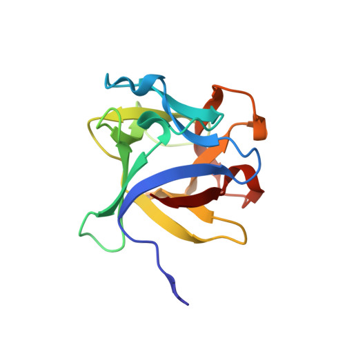
Structural analysis of the Rhizoctonia solani agglutinin reveals a domain-swapping dimeric assembly.

Skamnaki, V.T.Peumans, W.J.Kantsadi, A.L.Cubeta, M.A.Plas, K.Pakala, S.Zographos, S.E.Smagghe, G.Nierman, W.C.Van Damme, E.J.Leonidas, D.D.

(2013) FEBS J 280: 1750-1763

  • DOI: https://doi.org/10.1111/febs.12190
  • Primary Citation of Related Structures:  
    4G9M, 4G9N

  • PubMed Abstract: 

    Rhizoctonia solani agglutinin (RSA) is a 15.5-kDa lectin accumulated in the mycelium and sclerotia of the soil born plant pathogenic fungus R. solani. Although it is considered to serve as a storage protein and is implicated in fungal insecticidal activity, its physiological role remains unclear as a result of a lack of any structure/function relationship information. Glycan arrays showed that RSA displays high selectivity towards terminal nonreducing N-acetylgalactosamine residues. We determined the amino acid sequence of RSA and also determined the crystal structures of the free form and the RSA-N-acetylgalactosamine complex at 1.6 and 2.2 Å resolution, respectively. RSA is a homodimer comprised of two monomers adopting the β-trefoil fold. Each monomer accommodates two different carbohydrate-binding sites in an asymmetric way. Despite RSA topology similarities with R-type lectins, the two-monomer assembly involves an N-terminal swap, thus creating a dimer association novel to R-type lectins. Structural characterization of the two carbohydrate-binding sites offers insights on the structural determinants of the RSA carbohydrate specificity.


  • Organizational Affiliation

    Department of Biochemistry and Biotechnology, University of Thessaly, Larissa, Greece.


Macromolecules
Find similar proteins by:  (by identity cutoff)  |  3D Structure
Entity ID: 1
MoleculeChains Sequence LengthOrganismDetailsImage
agglutinin
A, B
143Rhizoctonia solaniMutation(s): 0 
UniProt
Find proteins for L8WGI4 (Thanatephorus cucumeris (strain AG1-IA))
Explore L8WGI4 
Go to UniProtKB:  L8WGI4
Entity Groups  
Sequence Clusters30% Identity50% Identity70% Identity90% Identity95% Identity100% Identity
UniProt GroupL8WGI4
Sequence Annotations
Expand
  • Reference Sequence
Experimental Data & Validation

Experimental Data

  • Method: X-RAY DIFFRACTION
  • Resolution: 2.20 Å
  • R-Value Free: 0.232 
  • R-Value Work: 0.150 
  • R-Value Observed: 0.154 
  • Space Group: C 1 2 1
Unit Cell:
Length ( Å )Angle ( ˚ )
a = 130.583α = 90
b = 61.251β = 93.45
c = 32.839γ = 90
Software Package:
Software NamePurpose
PHENIXrefinement
REFMACrefinement
CrysalisProdata collection
CrysalisProdata reduction
SCALAdata scaling
REFMACphasing

Structure Validation

View Full Validation Report


Ligand Structure Quality Assessment 


Entry History 

Deposition Data

Revision History  (Full details and data files)

  • Version 1.0: 2013-06-05
    Type: Initial release
  • Version 1.1: 2017-11-15
    Changes: Refinement description
  • Version 1.2: 2019-07-17
    Changes: Data collection, Refinement description
  • Version 1.3: 2020-07-29
    Type: Remediation
    Reason: Carbohydrate remediation
    Changes: Data collection, Derived calculations, Structure summary
  • Version 1.4: 2023-09-13
    Changes: Data collection, Database references, Refinement description, Structure summary